 Movie
Movie Controller

[English] 日本語
 Yorodumi
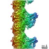
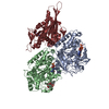
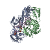
- PDB-6ta4: Human kinesin-5 motor domain in the AMPPNP state bound to microtubules -
+ Open data

- Basic information
| Entry | Database: PDB / ID: 6ta4 | |||||||||||||||
|---|---|---|---|---|---|---|---|---|---|---|---|---|---|---|---|---|
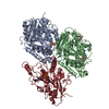
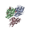
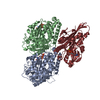
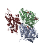
| Title | Human kinesin-5 motor domain in the AMPPNP state bound to microtubules | |||||||||||||||
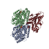
 Components Components |
| |||||||||||||||
 Keywords Keywords | CELL CYCLE / kinesin / microtubule / mitosis / inhibition / motor | |||||||||||||||
| Function / homology |  Function and homology information Function and homology informationspindle elongation / regulation of mitotic centrosome separation / plus-end-directed microtubule motor activity / Kinesins / Microtubule-dependent trafficking of connexons from Golgi to the plasma membrane / Resolution of Sister Chromatid Cohesion / Hedgehog 'off' state / Cilium Assembly / Intraflagellar transport / COPI-dependent Golgi-to-ER retrograde traffic ...spindle elongation / regulation of mitotic centrosome separation / plus-end-directed microtubule motor activity / Kinesins / Microtubule-dependent trafficking of connexons from Golgi to the plasma membrane / Resolution of Sister Chromatid Cohesion / Hedgehog 'off' state / Cilium Assembly / Intraflagellar transport / COPI-dependent Golgi-to-ER retrograde traffic / Mitotic Prometaphase / Carboxyterminal post-translational modifications of tubulin / RHOH GTPase cycle / EML4 and NUDC in mitotic spindle formation / Sealing of the nuclear envelope (NE) by ESCRT-III / Kinesins / PKR-mediated signaling / Separation of Sister Chromatids / The role of GTSE1 in G2/M progression after G2 checkpoint / Aggrephagy / RHO GTPases activate IQGAPs / RHO GTPases Activate Formins / HSP90 chaperone cycle for steroid hormone receptors (SHR) in the presence of ligand / spindle organization / MHC class II antigen presentation / Recruitment of NuMA to mitotic centrosomes / microtubule motor activity / kinesin complex / COPI-mediated anterograde transport / COPI-dependent Golgi-to-ER retrograde traffic / microtubule-based movement / mitotic spindle assembly / MHC class II antigen presentation / mitotic spindle organization / microtubule cytoskeleton organization / structural constituent of cytoskeleton / neuron migration / spindle / spindle pole / mitotic spindle / mitotic cell cycle / microtubule cytoskeleton / microtubule binding / microtubule / Hydrolases; Acting on acid anhydrides; Acting on GTP to facilitate cellular and subcellular movement / cell division / GTPase activity / protein kinase binding / GTP binding / protein-containing complex / ATP binding / membrane / metal ion binding / nucleus / cytosol / cytoplasm Similarity search - Function | |||||||||||||||
| Biological species |  Homo sapiens (human) Homo sapiens (human) | |||||||||||||||
| Method | ELECTRON MICROSCOPY / helical reconstruction / cryo EM / Resolution: 6.1 Å | |||||||||||||||
 Authors Authors | Pena, A.P. / Sweeney, A. / Cook, A.D. / Moores, C.A. / Topf, M. | |||||||||||||||
| Funding support |  United Kingdom, 4items United Kingdom, 4items
| |||||||||||||||
 Citation Citation | Journal: Structure / Year: 2020 Title: Structure of Microtubule-Trapped Human Kinesin-5 and Its Mechanism of Inhibition Revealed Using Cryoelectron Microscopy. Authors: Alejandro Peña / Aaron Sweeney / Alexander D Cook / Julia Locke / Maya Topf / Carolyn A Moores / Abstract: Kinesin-5 motors are vital mitotic spindle components, and disruption of their function perturbs cell division. We investigated the molecular mechanism of the human kinesin-5 inhibitor GSK-1, which ...Kinesin-5 motors are vital mitotic spindle components, and disruption of their function perturbs cell division. We investigated the molecular mechanism of the human kinesin-5 inhibitor GSK-1, which allosterically promotes tight microtubule binding. GSK-1 inhibits monomeric human kinesin-5 ATPase and microtubule gliding activities, and promotes the motor's microtubule stabilization activity. Using cryoelectron microscopy, we determined the 3D structure of the microtubule-bound motor-GSK-1 at 3.8 Å overall resolution. The structure reveals that GSK-1 stabilizes the microtubule binding surface of the motor in an ATP-like conformation, while destabilizing regions of the motor around the empty nucleotide binding pocket. Density corresponding to GSK-1 is located between helix-α4 and helix-α6 in the motor domain at its interface with the microtubule. Using a combination of difference mapping and protein-ligand docking, we characterized the kinesin-5-GSK-1 interaction and further validated this binding site using mutagenesis. This work opens up new avenues of investigation of kinesin inhibition and spindle perturbation. | |||||||||||||||
| History |
|
- Structure visualization
| Movie |
Movie viewer |
|---|---|
| Structure viewer | Molecule: MolmilJmol/JSmol |
- Downloads & links
-Download
| PDBx/mmCIF format | 6ta4.cif.gz | 234.7 KB | Display | PDBx/mmCIF format |
|---|---|---|---|---|
| PDB format | pdb6ta4.ent.gz | 183.5 KB | Display | PDB format |
| PDBx/mmJSON format | 6ta4.json.gz | Tree view | PDBx/mmJSON format | |
| Others |  Other downloads Other downloads |
-Validation report
| Arichive directory | https://data.pdbj.org/pub/pdb/validation_reports/ta/6ta4ftp://data.pdbj.org/pub/pdb/validation_reports/ta/6ta4 | HTTPS FTP |
|---|
-Related structure data
| Related structure data | 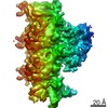 10422MC  6ta3C 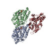 6tiwC M: map data used to model this data C: citing same article ( |
|---|---|
| Similar structure data |


-Links
 PDBj
PDBj


- Assembly
| Deposited unit | 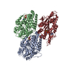
|
|---|---|
| 1 |
|
-Components
-Protein , 3 types, 3 molecules BAK
| #1: Protein | Mass: 48113.129 Da / Num. of mol.: 1 / Source method: isolated from a natural source / Source: (natural) |
|---|---|
| #2: Protein | Mass: 48780.117 Da / Num. of mol.: 1 / Source method: isolated from a natural source / Source: (natural) |
| #3: Protein | Mass: 43727.461 Da / Num. of mol.: 1 Source method: isolated from a genetically manipulated source Source: (gene. exp.) Homo sapiens (human) / Gene: KIF11, EG5, KNSL1, TRIP5 / Production host:  |
-Non-polymers , 4 types, 6 molecules 


| #4: Chemical | ChemComp-G2P /  Mass: 521.208 Da / Num. of mol.: 1 / Source method: obtained synthetically / Formula: C11H18N5O13P3 / Comment: GMP-CPP, energy-carrying molecule analogue*YM Mass: 521.208 Da / Num. of mol.: 1 / Source method: obtained synthetically / Formula: C11H18N5O13P3 / Comment: GMP-CPP, energy-carrying molecule analogue*YM | ||||
|---|---|---|---|---|---|
| #5: Chemical |  Mass: 24.305 Da / Num. of mol.: 3 / Source method: obtained synthetically / Formula: Mg Mass: 24.305 Da / Num. of mol.: 3 / Source method: obtained synthetically / Formula: Mg#6: Chemical | ChemComp-GTP / |  Mass: 523.180 Da / Num. of mol.: 1 / Source method: obtained synthetically / Formula: C10H16N5O14P3 / Comment: GTP, energy-carrying molecule*YM Mass: 523.180 Da / Num. of mol.: 1 / Source method: obtained synthetically / Formula: C10H16N5O14P3 / Comment: GTP, energy-carrying molecule*YM#7: Chemical | ChemComp-ANP / |  Mass: 506.196 Da / Num. of mol.: 1 / Source method: obtained synthetically / Formula: C10H17N6O12P3 / Feature type: SUBJECT OF INVESTIGATION / Comment: AMP-PNP, energy-carrying molecule analogue*YM Mass: 506.196 Da / Num. of mol.: 1 / Source method: obtained synthetically / Formula: C10H17N6O12P3 / Feature type: SUBJECT OF INVESTIGATION / Comment: AMP-PNP, energy-carrying molecule analogue*YM |
-Details
| Has ligand of interest | Y |
|---|
-Experimental details
-Experiment
| Experiment | Method: ELECTRON MICROSCOPY |
|---|---|
| EM experiment | Aggregation state: FILAMENT / 3D reconstruction method: helical reconstruction |
- Sample preparation
| Component |
| ||||||||||||||||||||||||
|---|---|---|---|---|---|---|---|---|---|---|---|---|---|---|---|---|---|---|---|---|---|---|---|---|---|
| Molecular weight | Experimental value: NO | ||||||||||||||||||||||||
| Source (natural) |
| ||||||||||||||||||||||||
| Source (recombinant) | Organism: | ||||||||||||||||||||||||
| Buffer solution | pH: 7.2 | ||||||||||||||||||||||||
| Specimen | Embedding applied: NO / Shadowing applied: NO / Staining applied: NO / Vitrification applied: YES | ||||||||||||||||||||||||
| Vitrification | Cryogen name: ETHANE |
- Electron microscopy imaging
| Experimental equipment |  Model: Tecnai Polara / Image courtesy: FEI Company |
|---|---|
| Microscopy | Model: FEI POLARA 300 |
| Electron gun | Electron source:  FIELD EMISSION GUN / Accelerating voltage: 300 kV / Illumination mode: FLOOD BEAM FIELD EMISSION GUN / Accelerating voltage: 300 kV / Illumination mode: FLOOD BEAM |
| Electron lens | Mode: BRIGHT FIELD |
| Image recording | Electron dose: 45 e/Å2 / Film or detector model: GATAN K2 SUMMIT (4k x 4k) |
- Processing
| CTF correction | Type: PHASE FLIPPING ONLY |
|---|---|
| Helical symmerty | Angular rotation/subunit: -25.7 ° / Axial rise/subunit: 8.95 Å / Axial symmetry: C1 |
| 3D reconstruction | Resolution: 6.1 Å / Resolution method: FSC 0.143 CUT-OFF / Num. of particles: 43714 / Symmetry type: HELICAL |
