 Movie
Movie Controller

[English] 日本語
 Yorodumi
- EMDB-10422: Human kinesin-5 motor domain in the AMPPNP state bound to microtubules -
+ Open data

- Basic information
| Entry | Database: EMDB / ID: EMD-10422 | |||||||||||||||
|---|---|---|---|---|---|---|---|---|---|---|---|---|---|---|---|---|
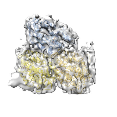
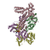
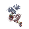
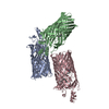
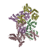
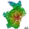
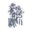
| Title | Human kinesin-5 motor domain in the AMPPNP state bound to microtubules | |||||||||||||||
 Map data Map data | ||||||||||||||||
 Sample Sample |
| |||||||||||||||
 Keywords Keywords | kinesin / microtubule / mitosis / inhibition / motor / CELL CYCLE | |||||||||||||||
| Function / homology |  Function and homology information Function and homology informationspindle elongation / regulation of mitotic centrosome separation / plus-end-directed microtubule motor activity / mitotic centrosome separation / Kinesins / Microtubule-dependent trafficking of connexons from Golgi to the plasma membrane / Resolution of Sister Chromatid Cohesion / Hedgehog 'off' state / Cilium Assembly / Intraflagellar transport ...spindle elongation / regulation of mitotic centrosome separation / plus-end-directed microtubule motor activity / mitotic centrosome separation / Kinesins / Microtubule-dependent trafficking of connexons from Golgi to the plasma membrane / Resolution of Sister Chromatid Cohesion / Hedgehog 'off' state / Cilium Assembly / Intraflagellar transport / COPI-dependent Golgi-to-ER retrograde traffic / Mitotic Prometaphase / Carboxyterminal post-translational modifications of tubulin / RHOH GTPase cycle / EML4 and NUDC in mitotic spindle formation / Sealing of the nuclear envelope (NE) by ESCRT-III / Kinesins / PKR-mediated signaling / Separation of Sister Chromatids / The role of GTSE1 in G2/M progression after G2 checkpoint / Aggrephagy / RHO GTPases activate IQGAPs / RHO GTPases Activate Formins / HSP90 chaperone cycle for steroid hormone receptors (SHR) in the presence of ligand / spindle organization / MHC class II antigen presentation / Recruitment of NuMA to mitotic centrosomes / microtubule motor activity / kinesin complex / COPI-mediated anterograde transport / COPI-dependent Golgi-to-ER retrograde traffic / microtubule-based movement / mitotic spindle assembly / MHC class II antigen presentation / mitotic spindle organization / sperm end piece / structural constituent of cytoskeleton / microtubule cytoskeleton organization / spindle / neuron migration / spindle pole / mitotic spindle / mitotic cell cycle / sperm principal piece / microtubule cytoskeleton / sperm midpiece / microtubule binding / microtubule / Hydrolases; Acting on acid anhydrides; Acting on GTP to facilitate cellular and subcellular movement / ciliary basal body / cell division / GTPase activity / protein kinase binding / GTP binding / protein-containing complex / ATP binding / membrane / metal ion binding / nucleus / cytoplasm / cytosol Similarity search - Function | |||||||||||||||
| Biological species |   Homo sapiens (human) Homo sapiens (human) | |||||||||||||||
| Method | helical reconstruction / cryo EM / Resolution: 6.1 Å | |||||||||||||||
 Authors Authors | Pena AP / Sweeney A | |||||||||||||||
| Funding support |  United Kingdom, 4 items United Kingdom, 4 items
| |||||||||||||||
 Citation Citation | Journal: Structure / Year: 2020 Title: Structure of Microtubule-Trapped Human Kinesin-5 and Its Mechanism of Inhibition Revealed Using Cryoelectron Microscopy. Authors: Alejandro Peña / Aaron Sweeney / Alexander D Cook / Julia Locke / Maya Topf / Carolyn A Moores / Abstract: Kinesin-5 motors are vital mitotic spindle components, and disruption of their function perturbs cell division. We investigated the molecular mechanism of the human kinesin-5 inhibitor GSK-1, which ...Kinesin-5 motors are vital mitotic spindle components, and disruption of their function perturbs cell division. We investigated the molecular mechanism of the human kinesin-5 inhibitor GSK-1, which allosterically promotes tight microtubule binding. GSK-1 inhibits monomeric human kinesin-5 ATPase and microtubule gliding activities, and promotes the motor's microtubule stabilization activity. Using cryoelectron microscopy, we determined the 3D structure of the microtubule-bound motor-GSK-1 at 3.8 Å overall resolution. The structure reveals that GSK-1 stabilizes the microtubule binding surface of the motor in an ATP-like conformation, while destabilizing regions of the motor around the empty nucleotide binding pocket. Density corresponding to GSK-1 is located between helix-α4 and helix-α6 in the motor domain at its interface with the microtubule. Using a combination of difference mapping and protein-ligand docking, we characterized the kinesin-5-GSK-1 interaction and further validated this binding site using mutagenesis. This work opens up new avenues of investigation of kinesin inhibition and spindle perturbation. | |||||||||||||||
| History |
|
- Structure visualization
| Movie |
Movie viewer |
|---|---|
| Structure viewer | EM map: SurfViewMolmilJmol/JSmol |
| Supplemental images |
- Downloads & links
-EMDB archive
| Map data | emd_10422.map.gz | 460.2 KB | EMDB map data format | |
|---|---|---|---|---|
| Header (meta data) | emd-10422-v30.xmlemd-10422.xml | 14.9 KB 14.9 KB | Display Display | EMDB header |
| FSC (resolution estimation) | emd_10422_fsc.xml | 13.9 KB | Display | FSC data file |
| Images |  emd_10422.png emd_10422.png | 159.8 KB | ||
| Filedesc metadata | emd-10422.cif.gz | 6.4 KB | ||
| Archive directory |  http://ftp.pdbj.org/pub/emdb/structures/EMD-10422ftp://ftp.pdbj.org/pub/emdb/structures/EMD-10422 http://ftp.pdbj.org/pub/emdb/structures/EMD-10422ftp://ftp.pdbj.org/pub/emdb/structures/EMD-10422 | HTTPS FTP |
-Related structure data
| Related structure data |  6ta4MC  6ta3C  6tiwC M: atomic model generated by this map C: citing same article ( |
|---|---|
| Similar structure data |


-Links
| EMDB pages | EMDB (EBI/PDBe) / EMDataResource |
|---|---|
| Related items in Molecule of the Month |


-Map
| File | Download / File: emd_10422.map.gz / Format: CCP4 / Size: 2.5 MB / Type: IMAGE STORED AS FLOATING POINT NUMBER (4 BYTES) | ||||||||||||||||||||||||||||||||||||||||||||||||||||||||||||
|---|---|---|---|---|---|---|---|---|---|---|---|---|---|---|---|---|---|---|---|---|---|---|---|---|---|---|---|---|---|---|---|---|---|---|---|---|---|---|---|---|---|---|---|---|---|---|---|---|---|---|---|---|---|---|---|---|---|---|---|---|---|
| Projections & slices | Image control
Images are generated by Spider. generated in cubic-lattice coordinate | ||||||||||||||||||||||||||||||||||||||||||||||||||||||||||||
| Voxel size | X=Y=Z: 1.39 Å | ||||||||||||||||||||||||||||||||||||||||||||||||||||||||||||
| Density |
| ||||||||||||||||||||||||||||||||||||||||||||||||||||||||||||
| Symmetry | Space group: 1 | ||||||||||||||||||||||||||||||||||||||||||||||||||||||||||||
| Details | EMDB XML:
CCP4 map header:
| ||||||||||||||||||||||||||||||||||||||||||||||||||||||||||||
 Z (Sec.)
 Y (Row.)
 X (Col.)


-Supplemental data
- Sample components
+Entire : AMPPNP bound Human Eg5 motor domain in complex with alpha/beta tubulin
+Supramolecule #1: AMPPNP bound Human Eg5 motor domain in complex with alpha/beta tubulin
+Supramolecule #2: AMPPNP bound Human Eg5 motor domain in complex with alpha/beta tubulin
+Supramolecule #3: AMPPNP bound Human Eg5 motor domain in complex with alpha/beta tubulin
+Macromolecule #1: Tubulin beta chain
+Macromolecule #2: Tubulin alpha-1B chain
+Macromolecule #3: Kinesin-like protein KIF11

+Macromolecule #4: PHOSPHOMETHYLPHOSPHONIC ACID GUANYLATE ESTER

+Macromolecule #5: MAGNESIUM ION
+Macromolecule #6: GUANOSINE-5'-TRIPHOSPHATE

+Macromolecule #7: PHOSPHOAMINOPHOSPHONIC ACID-ADENYLATE ESTER

-Experimental details
-Structure determination
| Method | cryo EM |
|---|---|
 Processing Processing | helical reconstruction |
| Aggregation state | filament |
-Sample preparation
| Buffer | pH: 7.2 |
|---|---|
| Vitrification | Cryogen name: ETHANE |
- Electron microscopy
| Microscope | FEI POLARA 300 |
|---|---|
| Image recording | Film or detector model: GATAN K2 SUMMIT (4k x 4k) / Average electron dose: 45.0 e/Å2 |
| Electron beam | Acceleration voltage: 300 kV / Electron source:  FIELD EMISSION GUN FIELD EMISSION GUN |
| Electron optics | Illumination mode: FLOOD BEAM / Imaging mode: BRIGHT FIELD |
| Experimental equipment |  Model: Tecnai Polara / Image courtesy: FEI Company |
-Image processing
| Final reconstruction | Applied symmetry - Helical parameters - Δz: 8.95 Å Applied symmetry - Helical parameters - Δ&Phi: -25.7 ° Applied symmetry - Helical parameters - Axial symmetry: C1 (asymmetric) Resolution.type: BY AUTHOR / Resolution: 6.1 Å / Resolution method: FSC 0.143 CUT-OFF / Number images used: 43714 |
|---|---|
| Startup model | Type of model: INSILICO MODEL |
| Final angle assignment | Type: NOT APPLICABLE |
| FSC plot (resolution estimation) | 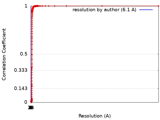 |
