 Movie
Movie Controller

[English] 日本語
 Yorodumi
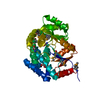
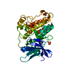
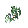
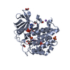
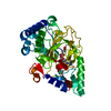
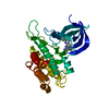
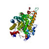
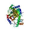
- PDB-6t6d: Crystal structure of the ACVR1 (ALK2) kinase in complex with the ... -
+ Open data

- Basic information
| Entry | Database: PDB / ID: 6t6d | ||||||
|---|---|---|---|---|---|---|---|
| Title | Crystal structure of the ACVR1 (ALK2) kinase in complex with the compound M4K2149 | ||||||
 Components Components | Activin receptor type I | ||||||
 Keywords Keywords | SIGNALING PROTEIN / KINASE / BMP / INHIBITOR / SIGNALLING | ||||||
| Function / homology |  Function and homology information Function and homology informationendocardial cushion cell fate commitment / acute inflammatory response / mitral valve morphogenesis / BMP receptor complex / BMP receptor activity / endocardial cushion fusion / cardiac muscle cell fate commitment / atrial septum primum morphogenesis / positive regulation of cardiac epithelial to mesenchymal transition / positive regulation of determination of dorsal identity ...endocardial cushion cell fate commitment / acute inflammatory response / mitral valve morphogenesis / BMP receptor complex / BMP receptor activity / endocardial cushion fusion / cardiac muscle cell fate commitment / atrial septum primum morphogenesis / positive regulation of cardiac epithelial to mesenchymal transition / positive regulation of determination of dorsal identity / transforming growth factor beta receptor activity, type I / smooth muscle cell differentiation / activin receptor activity, type I / activin receptor complex / pharyngeal system development / endocardial cushion formation / receptor protein serine/threonine kinase / activin binding / cellular response to BMP stimulus / transmembrane receptor protein serine/threonine kinase activity / activin receptor signaling pathway / negative regulation of activin receptor signaling pathway / gastrulation with mouth forming second / embryonic heart tube morphogenesis / dorsal/ventral pattern formation / transforming growth factor beta binding / determination of left/right symmetry / atrioventricular valve morphogenesis / neural crest cell migration / branching involved in blood vessel morphogenesis / ventricular septum morphogenesis / negative regulation of G1/S transition of mitotic cell cycle / SMAD binding / germ cell development / mesoderm formation / peptide hormone binding / positive regulation of SMAD protein signal transduction / positive regulation of intracellular signal transduction / regulation of ossification / positive regulation of osteoblast differentiation / BMP signaling pathway / negative regulation of signal transduction / positive regulation of bone mineralization / transforming growth factor beta receptor signaling pathway / protein tyrosine kinase binding / negative regulation of extrinsic apoptotic signaling pathway / cellular response to growth factor stimulus / osteoblast differentiation / apical part of cell / heart development / in utero embryonic development / protein kinase activity / cell differentiation / positive regulation of cell migration / cadherin binding / protein serine/threonine kinase activity / positive regulation of DNA-templated transcription / protein homodimerization activity / positive regulation of transcription by RNA polymerase II / ATP binding / metal ion binding / plasma membrane Similarity search - Function | ||||||
| Biological species |  Homo sapiens (human) Homo sapiens (human) | ||||||
| Method |  X-RAY DIFFRACTION / SYNCHROTRON / MOLECULAR REPLACEMENT / Resolution: 2.56 Å X-RAY DIFFRACTION / SYNCHROTRON / MOLECULAR REPLACEMENT / Resolution: 2.56 Å | ||||||
 Authors Authors | Adamson, R.J. / Williams, E.P. / Smil, D. / Burgess-Brown, N. / von Delft, F. / Arrowsmith, C.H. / Edwards, A.M. / Bountra, C. / Bullock, A.N. | ||||||
 Citation Citation | Journal: J.Med.Chem. / Year: 2020 Title: Targeting ALK2: An Open Science Approach to Developing Therapeutics for the Treatment of Diffuse Intrinsic Pontine Glioma. Authors: Ensan, D. / Smil, D. / Zepeda-Velazquez, C.A. / Panagopoulos, D. / Wong, J.F. / Williams, E.P. / Adamson, R. / Bullock, A.N. / Kiyota, T. / Aman, A. / Roberts, O.G. / Edwards, A.M. / ...Authors: Ensan, D. / Smil, D. / Zepeda-Velazquez, C.A. / Panagopoulos, D. / Wong, J.F. / Williams, E.P. / Adamson, R. / Bullock, A.N. / Kiyota, T. / Aman, A. / Roberts, O.G. / Edwards, A.M. / O'Meara, J.A. / Isaac, M.B. / Al-Awar, R. | ||||||
| History |
|
- Structure visualization
| Structure viewer | Molecule: MolmilJmol/JSmol |
|---|
- Downloads & links
-Download
| PDBx/mmCIF format | 6t6d.cif.gz | 294.6 KB | Display | PDBx/mmCIF format |
|---|---|---|---|---|
| PDB format | pdb6t6d.ent.gz | 192.7 KB | Display | PDB format |
| PDBx/mmJSON format | 6t6d.json.gz | Tree view | PDBx/mmJSON format | |
| Others |  Other downloads Other downloads |
-Validation report
| Arichive directory | https://data.pdbj.org/pub/pdb/validation_reports/t6/6t6dftp://data.pdbj.org/pub/pdb/validation_reports/t6/6t6d | HTTPS FTP |
|---|
-Related structure data
| Related structure data |  8r7gC  6srhS S: Starting model for refinement C: citing same article ( |
|---|---|
| Similar structure data |


-Links
 PDBj
PDBj
- Assembly
| Deposited unit | 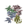
| ||||||||||||
|---|---|---|---|---|---|---|---|---|---|---|---|---|---|
| 1 | 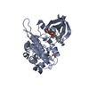
| ||||||||||||
| 2 | 
| ||||||||||||
| 3 | 
| ||||||||||||
| 4 | 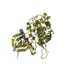
| ||||||||||||
| Unit cell |
|
-Components
| #1: Protein | Mass: 34537.633 Da / Num. of mol.: 4 / Mutation: Q207D Source method: isolated from a genetically manipulated source Details: Chains E, F, G and H contain ligand 149 bound to the kinase active site and SO4 as part of the crystal packing. Source: (gene. exp.) Homo sapiens (human) / Gene: ACVR1 / Cell line (production host): Sf9 / Production host:   Spodoptera frugiperda (fall armyworm) Spodoptera frugiperda (fall armyworm)References: UniProt: Q04771, receptor protein serine/threonine kinase #2: Chemical | ChemComp-MM8 /   Mass: 402.489 Da / Num. of mol.: 4 / Source method: obtained synthetically / Formula: C24H26N4O2 / Feature type: SUBJECT OF INVESTIGATION Mass: 402.489 Da / Num. of mol.: 4 / Source method: obtained synthetically / Formula: C24H26N4O2 / Feature type: SUBJECT OF INVESTIGATION#3: Chemical | ChemComp-SO4 /   Mass: 96.063 Da / Num. of mol.: 11 / Source method: isolated from a natural source / Formula: SO4 Mass: 96.063 Da / Num. of mol.: 11 / Source method: isolated from a natural source / Formula: SO4#4: Water | ChemComp-HOH / |  Mass: 18.015 Da / Num. of mol.: 30 / Source method: isolated from a natural source / Formula: H2O Mass: 18.015 Da / Num. of mol.: 30 / Source method: isolated from a natural source / Formula: H2OHas ligand of interest | Y | Has protein modification | Y | |
|---|
-Experimental details
-Experiment
| Experiment | Method: X-RAY DIFFRACTION / Number of used crystals: 1 |
|---|
- Sample preparation
| Crystal | Density Matthews: 2.92 Å3/Da / Density % sol: 57.9 % |
|---|---|
| Crystal grow | Temperature: 277 K / Method: vapor diffusion, sitting drop / pH: 4.9 Details: 0.1M citrate pH 4.9, 1M ammonium sulfate, 0.2M sodium/potassium tartrate |
-Data collection
| Diffraction | Mean temperature: 100 K / Serial crystal experiment: N |
|---|---|
| Diffraction source | Source: SYNCHROTRON / Site: Diamond  / Beamline: I03 / Wavelength: 0.9763 Å / Beamline: I03 / Wavelength: 0.9763 Å |
| Detector | Type: ADSC QUANTUM 315 / Detector: CCD / Date: Jan 21, 2019 |
| Radiation | Monochromator: double crystal / Protocol: SINGLE WAVELENGTH / Monochromatic (M) / Laue (L): M / Scattering type: x-ray |
| Radiation wavelength | Wavelength: 0.9763 Å / Relative weight: 1 |
| Reflection | Resolution: 2.56→89.13 Å / Num. obs: 50340 / % possible obs: 100 % / Redundancy: 13.3 % / Biso Wilson estimate: 58.84 Å2 / CC1/2: 0.998 / Rmerge(I) obs: 0.197 / Rpim(I) all: 0.056 / Rrim(I) all: 0.205 / Χ2: 0.99 / Net I/σ(I): 8.8 |
| Reflection shell | Resolution: 2.56→2.63 Å / Redundancy: 13.5 % / Rmerge(I) obs: 3.197 / Mean I/σ(I) obs: 1.3 / Num. unique obs: 3699 / CC1/2: 0.87 / Rpim(I) all: 0.901 / Rrim(I) all: 3.323 / Χ2: 1 / % possible all: 100 |
- Processing
| Software |
| |||||||||||||||||||||||||||||||||||||||||||||||||||||||||||||||||||||||||||||||||||||||||||||||||||||||||||||||||||||||||||||||||||||
|---|---|---|---|---|---|---|---|---|---|---|---|---|---|---|---|---|---|---|---|---|---|---|---|---|---|---|---|---|---|---|---|---|---|---|---|---|---|---|---|---|---|---|---|---|---|---|---|---|---|---|---|---|---|---|---|---|---|---|---|---|---|---|---|---|---|---|---|---|---|---|---|---|---|---|---|---|---|---|---|---|---|---|---|---|---|---|---|---|---|---|---|---|---|---|---|---|---|---|---|---|---|---|---|---|---|---|---|---|---|---|---|---|---|---|---|---|---|---|---|---|---|---|---|---|---|---|---|---|---|---|---|---|---|---|
| Refinement | Method to determine structure: MOLECULAR REPLACEMENT Starting model: 6SRH Resolution: 2.56→78.45 Å / SU ML: 0.4258 / Cross valid method: FREE R-VALUE / σ(F): 1.33 / Phase error: 35.6209
| |||||||||||||||||||||||||||||||||||||||||||||||||||||||||||||||||||||||||||||||||||||||||||||||||||||||||||||||||||||||||||||||||||||
| Solvent computation | Shrinkage radii: 0.9 Å / VDW probe radii: 1.11 Å | |||||||||||||||||||||||||||||||||||||||||||||||||||||||||||||||||||||||||||||||||||||||||||||||||||||||||||||||||||||||||||||||||||||
| Displacement parameters | Biso mean: 73.66 Å2 | |||||||||||||||||||||||||||||||||||||||||||||||||||||||||||||||||||||||||||||||||||||||||||||||||||||||||||||||||||||||||||||||||||||
| Refinement step | Cycle: LAST / Resolution: 2.56→78.45 Å
| |||||||||||||||||||||||||||||||||||||||||||||||||||||||||||||||||||||||||||||||||||||||||||||||||||||||||||||||||||||||||||||||||||||
| Refine LS restraints |
| |||||||||||||||||||||||||||||||||||||||||||||||||||||||||||||||||||||||||||||||||||||||||||||||||||||||||||||||||||||||||||||||||||||
| LS refinement shell |
|
