 Movie
Movie Controller

[English] 日本語
 Yorodumi
- PDB-6ssp: Structure of the pentameric ligand-gated ion channel ELIC in comp... -
+ Open data

- Basic information
| Entry | Database: PDB / ID: 6ssp | ||||||||||||||||||
|---|---|---|---|---|---|---|---|---|---|---|---|---|---|---|---|---|---|---|---|
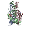
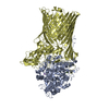
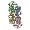
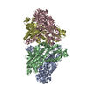
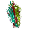
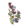
| Title | Structure of the pentameric ligand-gated ion channel ELIC in complex with a NAM nanobody | ||||||||||||||||||
 Components Components |
| ||||||||||||||||||
 Keywords Keywords | TRANSPORT PROTEIN / ELIC / CYS-LOOP RECEPTOR / NANOBODY / PENTAMERIC LIGAND GATED ION CHANNEL | ||||||||||||||||||
| Function / homology |  Function and homology information Function and homology informationextracellular ligand-gated monoatomic ion channel activity / transmembrane signaling receptor activity / identical protein binding / plasma membrane Similarity search - Function | ||||||||||||||||||
| Biological species |  Dickeya chrysanthemi (bacteria) Dickeya chrysanthemi (bacteria) | ||||||||||||||||||
| Method |  X-RAY DIFFRACTION / SYNCHROTRON / MOLECULAR REPLACEMENT / Resolution: 3.25 Å X-RAY DIFFRACTION / SYNCHROTRON / MOLECULAR REPLACEMENT / Resolution: 3.25 Å | ||||||||||||||||||
 Authors Authors | Ulens, C. / Brams, M. / Evans, G.L. / Spurny, R. / Govaerts, C. / Pardon, E. / Steyaert, J. | ||||||||||||||||||
| Funding support |  Belgium, 5items Belgium, 5items
| ||||||||||||||||||
 Citation Citation | Journal: Elife / Year: 2020 Title: Modulation of the Erwinia ligand-gated ion channel (ELIC) and the 5-HT 3 receptor via a common vestibule site. Authors: Brams, M. / Govaerts, C. / Kambara, K. / Price, K.L. / Spurny, R. / Gharpure, A. / Pardon, E. / Evans, G.L. / Bertrand, D. / Lummis, S.C. / Hibbs, R.E. / Steyaert, J. / Ulens, C. | ||||||||||||||||||
| History |
|
- Structure visualization
| Structure viewer | Molecule: MolmilJmol/JSmol |
|---|
- Downloads & links
-Download
| PDBx/mmCIF format | 6ssp.cif.gz | 651.2 KB | Display | PDBx/mmCIF format |
|---|---|---|---|---|
| PDB format | pdb6ssp.ent.gz | 530.9 KB | Display | PDB format |
| PDBx/mmJSON format | 6ssp.json.gz | Tree view | PDBx/mmJSON format | |
| Others |  Other downloads Other downloads |
-Validation report
| Arichive directory | https://data.pdbj.org/pub/pdb/validation_reports/ss/6sspftp://data.pdbj.org/pub/pdb/validation_reports/ss/6ssp | HTTPS FTP |
|---|
-Related structure data
| Related structure data |  6ssiC  2vl0S 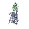 3p0gS C: citing same article ( S: Starting model for refinement |
|---|---|
| Similar structure data |


-Links
 PDBj
PDBj


- Assembly
| Deposited unit | 
| ||||||||
|---|---|---|---|---|---|---|---|---|---|
| 1 | 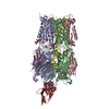
| ||||||||
| 2 | 
| ||||||||
| Unit cell |
|
-Components
| #1: Protein | Mass: 36465.578 Da / Num. of mol.: 10 Source method: isolated from a genetically manipulated source Source: (gene. exp.) Dickeya chrysanthemi (bacteria) / Production host: #2: Antibody | Mass: 15250.717 Da / Num. of mol.: 2 Source method: isolated from a genetically manipulated source Source: (gene. exp.) #3: Chemical | ChemComp-CA /   Mass: 40.078 Da / Num. of mol.: 8 / Source method: obtained synthetically / Formula: Ca Mass: 40.078 Da / Num. of mol.: 8 / Source method: obtained synthetically / Formula: Ca#4: Chemical | ChemComp-UMQ /   Mass: 496.589 Da / Num. of mol.: 10 / Source method: obtained synthetically / Formula: C23H44O11 / Comment: detergent*YM Mass: 496.589 Da / Num. of mol.: 10 / Source method: obtained synthetically / Formula: C23H44O11 / Comment: detergent*YMHas ligand of interest | N | Has protein modification | Y | |
|---|
-Experimental details
-Experiment
| Experiment | Method: X-RAY DIFFRACTION / Number of used crystals: 1 |
|---|
- Sample preparation
| Crystal | Density Matthews: 3.99 Å3/Da / Density % sol: 69.19 % |
|---|---|
| Crystal grow | Temperature: 293 K / Method: vapor diffusion, sitting drop / pH: 8.5 / Details: 0.1 M Na2SO4, 0.1 M bis-trispropane, 10% PEG3350 |
-Data collection
| Diffraction | Mean temperature: 100 K / Serial crystal experiment: N | ||||||||||||||||||||||||||||||
|---|---|---|---|---|---|---|---|---|---|---|---|---|---|---|---|---|---|---|---|---|---|---|---|---|---|---|---|---|---|---|---|
| Diffraction source | Source: SYNCHROTRON / Site: SLS  / Beamline: X06DA / Wavelength: 1 Å / Beamline: X06DA / Wavelength: 1 Å | ||||||||||||||||||||||||||||||
| Detector | Type: DECTRIS PILATUS 2M-F / Detector: PIXEL / Date: Nov 26, 2011 | ||||||||||||||||||||||||||||||
| Radiation | Protocol: SINGLE WAVELENGTH / Monochromatic (M) / Laue (L): M / Scattering type: x-ray | ||||||||||||||||||||||||||||||
| Radiation wavelength | Wavelength: 1 Å / Relative weight: 1 | ||||||||||||||||||||||||||||||
| Reflection | Resolution: 3.25→48.32 Å / Num. obs: 82177 / % possible obs: 89.8 % / Redundancy: 1.8 % / Biso Wilson estimate: 105.94 Å2 / CC1/2: 0.993 / Rmerge(I) obs: 0.053 / Rpim(I) all: 0.053 / Rrim(I) all: 0.074 / Net I/σ(I): 5.6 / Num. measured all: 146197 | ||||||||||||||||||||||||||||||
| Reflection shell | Diffraction-ID: 1
|
- Processing
| Software |
| ||||||||||||||||||||||||||||||||||||||||||||||||||||||||||||||||||||||||||||||||||||||||||||||||||||||||||||
|---|---|---|---|---|---|---|---|---|---|---|---|---|---|---|---|---|---|---|---|---|---|---|---|---|---|---|---|---|---|---|---|---|---|---|---|---|---|---|---|---|---|---|---|---|---|---|---|---|---|---|---|---|---|---|---|---|---|---|---|---|---|---|---|---|---|---|---|---|---|---|---|---|---|---|---|---|---|---|---|---|---|---|---|---|---|---|---|---|---|---|---|---|---|---|---|---|---|---|---|---|---|---|---|---|---|---|---|---|---|
| Refinement | Method to determine structure: MOLECULAR REPLACEMENT Starting model: 2vl0, 3p0g Resolution: 3.25→48.32 Å / Cor.coef. Fo:Fc: 0.87 / Cor.coef. Fo:Fc free: 0.86 / Cross valid method: THROUGHOUT / σ(F): 0 / SU Rfree Blow DPI: 0.474
| ||||||||||||||||||||||||||||||||||||||||||||||||||||||||||||||||||||||||||||||||||||||||||||||||||||||||||||
| Displacement parameters | Biso max: 241.96 Å2 / Biso mean: 104.73 Å2 / Biso min: 46.74 Å2
| ||||||||||||||||||||||||||||||||||||||||||||||||||||||||||||||||||||||||||||||||||||||||||||||||||||||||||||
| Refine analyze | Luzzati coordinate error obs: 0.56 Å | ||||||||||||||||||||||||||||||||||||||||||||||||||||||||||||||||||||||||||||||||||||||||||||||||||||||||||||
| Refinement step | Cycle: final / Resolution: 3.25→48.32 Å
| ||||||||||||||||||||||||||||||||||||||||||||||||||||||||||||||||||||||||||||||||||||||||||||||||||||||||||||
| Refine LS restraints |
| ||||||||||||||||||||||||||||||||||||||||||||||||||||||||||||||||||||||||||||||||||||||||||||||||||||||||||||
| LS refinement shell | Resolution: 3.25→3.29 Å / Rfactor Rfree error: 0 / Total num. of bins used: 50
|
