 Movie
Movie Controller

[English] 日本語
 Yorodumi
- PDB-2vl0: X-ray structure of a pentameric ligand gated ion channel from Erw... -
+ Open data

- Basic information
| Entry | Database: PDB / ID: 2vl0 | ||||||
|---|---|---|---|---|---|---|---|
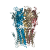
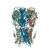
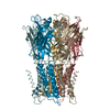
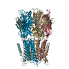
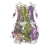
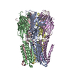
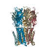
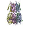
| Title | X-ray structure of a pentameric ligand gated ion channel from Erwinia chrysanthemi (ELIC) | ||||||
 Components Components | Cys-loop ligand-gated ion channel | ||||||
 Keywords Keywords | MEMBRANE PROTEIN / PENTAMERIC LIGAND GATED ION CHANNEL / PROKARYOTIC CYS-LOOP RECEPTOR / CATION SELECTIVE CHANNEL | ||||||
| Function / homology |  Function and homology information Function and homology informationextracellular ligand-gated monoatomic ion channel activity / transmembrane signaling receptor activity / identical protein binding / plasma membrane Similarity search - Function | ||||||
| Biological species |  Dickeya chrysanthemi (bacteria) Dickeya chrysanthemi (bacteria) | ||||||
| Method |  X-RAY DIFFRACTION / SYNCHROTRON / SIRAS / Resolution: 3.3 Å X-RAY DIFFRACTION / SYNCHROTRON / SIRAS / Resolution: 3.3 Å | ||||||
 Authors Authors | Hilf, R.J.C. / Dutzler, R. | ||||||
 Citation Citation | Journal: Nature / Year: 2008 Title: X-Ray Structure of a Prokaryotic Pentameric Ligand-Gated Ion Channel Authors: Hilf, R.J.C. / Dutzler, R. | ||||||
| History |
| ||||||
| Remark 700 | SHEET THE SHEET STRUCTURE OF THIS MOLECULE IS BIFURCATED. IN ORDER TO REPRESENT THIS FEATURE IN ... SHEET THE SHEET STRUCTURE OF THIS MOLECULE IS BIFURCATED. IN ORDER TO REPRESENT THIS FEATURE IN THE SHEET RECORDS BELOW, TWO SHEETS ARE DEFINED. |
- Structure visualization
| Structure viewer | Molecule: MolmilJmol/JSmol |
|---|
- Downloads & links
-Download
| PDBx/mmCIF format | 2vl0.cif.gz | 602 KB | Display | PDBx/mmCIF format |
|---|---|---|---|---|
| PDB format | pdb2vl0.ent.gz | 499.8 KB | Display | PDB format |
| PDBx/mmJSON format | 2vl0.json.gz | Tree view | PDBx/mmJSON format | |
| Others |  Other downloads Other downloads |
-Validation report
| Arichive directory | https://data.pdbj.org/pub/pdb/validation_reports/vl/2vl0ftp://data.pdbj.org/pub/pdb/validation_reports/vl/2vl0 | HTTPS FTP |
|---|
-Related structure data
| Similar structure data |
|---|

-Links
 PDBj
PDBj

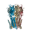
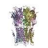
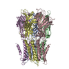
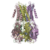
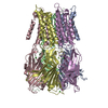
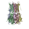
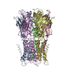
- Assembly
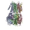
| Deposited unit | 
| ||||||||||||||||||||||||||||||||||||||||
|---|---|---|---|---|---|---|---|---|---|---|---|---|---|---|---|---|---|---|---|---|---|---|---|---|---|---|---|---|---|---|---|---|---|---|---|---|---|---|---|---|---|
| 1 | 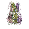
| ||||||||||||||||||||||||||||||||||||||||
| 2 | 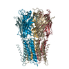
| ||||||||||||||||||||||||||||||||||||||||
| Unit cell |
| ||||||||||||||||||||||||||||||||||||||||
| Noncrystallographic symmetry (NCS) | NCS oper:
|
-Components
| #1: Protein | Mass: 36821.949 Da / Num. of mol.: 10 Source method: isolated from a genetically manipulated source Source: (gene. exp.) Dickeya chrysanthemi (bacteria) / Plasmid: PET26 DERIVATIVE / Production host: |
|---|
-Experimental details
-Experiment
| Experiment | Method: X-RAY DIFFRACTION / Number of used crystals: 1 |
|---|
- Sample preparation
| Crystal | Density Matthews: 4.19 Å3/Da / Density % sol: 65 % Description: PHASES WERE EXTENDED BY CYCLIC 10-FOLD NCS AVERAGING |
|---|---|
| Crystal grow | pH: 6.5 Details: 15 % PEG 4000, 200MM AMMONIUM SULFATE, 50 MM ADA PH 6.5 |
-Data collection
| Diffraction | Mean temperature: 100 K |
|---|---|
| Diffraction source | Source: SYNCHROTRON / Site: SLS  / Beamline: X06SA / Wavelength: 0.979 / Beamline: X06SA / Wavelength: 0.979 |
| Detector | Type: DECTRIS CUSTOM / Date: Sep 7, 2007 |
| Radiation | Protocol: SINGLE WAVELENGTH / Monochromatic (M) / Laue (L): M / Scattering type: x-ray |
| Radiation wavelength | Wavelength: 0.979 Å / Relative weight: 1 |
| Reflection | Resolution: 3.3→40 Å / Num. obs: 86136 / % possible obs: 99.7 % / Observed criterion σ(I): -3 / Redundancy: 21 % / Rmerge(I) obs: 0.14 / Net I/σ(I): 17.4 |
| Reflection shell | Resolution: 3.3→3.4 Å / Redundancy: 15.1 % / Rmerge(I) obs: 1.12 / Mean I/σ(I) obs: 2.7 / % possible all: 99.9 |
- Processing
| Software |
| ||||||||||||||||||||||||||||||||||||||||||||||||||||||||||||||||||||||||||||||||
|---|---|---|---|---|---|---|---|---|---|---|---|---|---|---|---|---|---|---|---|---|---|---|---|---|---|---|---|---|---|---|---|---|---|---|---|---|---|---|---|---|---|---|---|---|---|---|---|---|---|---|---|---|---|---|---|---|---|---|---|---|---|---|---|---|---|---|---|---|---|---|---|---|---|---|---|---|---|---|---|---|---|
| Refinement | Method to determine structure: SIRAS Starting model: NONE Resolution: 3.3→29.93 Å / Rfactor Rfree error: 0.004 / Data cutoff high absF: 4877940.57 / Isotropic thermal model: RESTRAINED / Cross valid method: THROUGHOUT / σ(F): 0 / Stereochemistry target values: MAXIMUM LIKELYHOOD / Details: BULK SOLVENT MODEL USED
| ||||||||||||||||||||||||||||||||||||||||||||||||||||||||||||||||||||||||||||||||
| Solvent computation | Solvent model: FLAT MODEL / Bsol: 61.2833 Å2 / ksol: 0.3 e/Å3 | ||||||||||||||||||||||||||||||||||||||||||||||||||||||||||||||||||||||||||||||||
| Displacement parameters | Biso mean: 129.1 Å2
| ||||||||||||||||||||||||||||||||||||||||||||||||||||||||||||||||||||||||||||||||
| Refine analyze |
| ||||||||||||||||||||||||||||||||||||||||||||||||||||||||||||||||||||||||||||||||
| Refinement step | Cycle: LAST / Resolution: 3.3→29.93 Å
| ||||||||||||||||||||||||||||||||||||||||||||||||||||||||||||||||||||||||||||||||
| Refine LS restraints |
| ||||||||||||||||||||||||||||||||||||||||||||||||||||||||||||||||||||||||||||||||
| Refine LS restraints NCS | NCS model details: TIGHT NCS POSITIONAL AND B-FACTOR RESTRAINTS Weight Biso : 5 / Weight position: 100 | ||||||||||||||||||||||||||||||||||||||||||||||||||||||||||||||||||||||||||||||||
| LS refinement shell | Resolution: 3.3→3.51 Å / Rfactor Rfree error: 0.017 / Total num. of bins used: 6
| ||||||||||||||||||||||||||||||||||||||||||||||||||||||||||||||||||||||||||||||||
| Xplor file | Serial no: 1 / Param file: PROTEIN_REP.PARAM / Topol file: PROTEIN.TOP |
