 Movie
Movie Controller

[English] 日本語
 Yorodumi
- PDB-6smw: A. thaliana serine hydroxymethyltransferase isoform 2 (AtSHMT2) i... -
+ Open data

- Basic information
| Entry | Database: PDB / ID: 6smw | ||||||
|---|---|---|---|---|---|---|---|
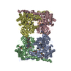
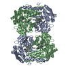
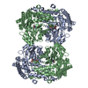
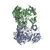
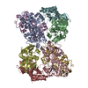
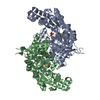
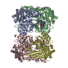
| Title | A. thaliana serine hydroxymethyltransferase isoform 2 (AtSHMT2) in complex with pemetrexed | ||||||
 Components Components | Serine hydroxymethyltransferase 2, mitochondrial | ||||||
 Keywords Keywords | TRANSFERASE / SHMT / antifolate | ||||||
| Function / homology |  Function and homology information Function and homology informationglycine metabolic process / L-serine metabolic process / glycine hydroxymethyltransferase / glycine hydroxymethyltransferase activity / : / tetrahydrofolate metabolic process / tetrahydrofolate interconversion / cobalt ion binding / plastid / respiratory chain complex I ...glycine metabolic process / L-serine metabolic process / glycine hydroxymethyltransferase / glycine hydroxymethyltransferase activity / : / tetrahydrofolate metabolic process / tetrahydrofolate interconversion / cobalt ion binding / plastid / respiratory chain complex I / chloroplast / pyridoxal phosphate binding / mitochondrion / zinc ion binding Similarity search - Function | ||||||
| Biological species |  | ||||||
| Method |  X-RAY DIFFRACTION / SYNCHROTRON / MOLECULAR REPLACEMENT / Resolution: 1.54 Å X-RAY DIFFRACTION / SYNCHROTRON / MOLECULAR REPLACEMENT / Resolution: 1.54 Å | ||||||
 Authors Authors | Ruszkowski, M. / Sekula, B. / Dauter, Z. | ||||||
 Citation Citation | Journal: Sci Rep / Year: 2019 Title: Structural basis of methotrexate and pemetrexed action on serine hydroxymethyltransferases revealed using plant models. Authors: Ruszkowski, M. / Sekula, B. / Ruszkowska, A. / Contestabile, R. / Nogues, I. / Angelaccio, S. / Szczepaniak, A. / Dauter, Z. | ||||||
| History |
|
- Structure visualization
| Structure viewer | Molecule: MolmilJmol/JSmol |
|---|
- Downloads & links
-Download
| PDBx/mmCIF format | 6smw.cif.gz | 862.3 KB | Display | PDBx/mmCIF format |
|---|---|---|---|---|
| PDB format | pdb6smw.ent.gz | 706 KB | Display | PDB format |
| PDBx/mmJSON format | 6smw.json.gz | Tree view | PDBx/mmJSON format | |
| Others |  Other downloads Other downloads |
-Validation report
| Arichive directory | https://data.pdbj.org/pub/pdb/validation_reports/sm/6smwftp://data.pdbj.org/pub/pdb/validation_reports/sm/6smw | HTTPS FTP |
|---|
-Related structure data
| Related structure data |  6smnC  6smrC  6cd0S S: Starting model for refinement C: citing same article ( |
|---|---|
| Similar structure data |


-Links
 PDBj
PDBj- Assembly
| Deposited unit | 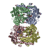
| ||||||||||||
|---|---|---|---|---|---|---|---|---|---|---|---|---|---|
| 1 |
| ||||||||||||
| Unit cell |
|
-Components
-Protein , 1 types, 4 molecules ABCD
| #1: Protein | Mass: 53545.035 Da / Num. of mol.: 4 Source method: isolated from a genetically manipulated source Source: (gene. exp.)  References: UniProt: Q94C74, glycine hydroxymethyltransferase |
|---|
-Non-polymers , 6 types, 1982 molecules 


| #2: Chemical |  Mass: 336.235 Da / Num. of mol.: 3 / Source method: obtained synthetically / Formula: C11H17N2O8P Mass: 336.235 Da / Num. of mol.: 3 / Source method: obtained synthetically / Formula: C11H17N2O8P#3: Chemical | ChemComp-EDO /  Mass: 62.068 Da / Num. of mol.: 38 / Source method: obtained synthetically / Formula: C2H6O2 Mass: 62.068 Da / Num. of mol.: 38 / Source method: obtained synthetically / Formula: C2H6O2#4: Chemical |  Mass: 427.411 Da / Num. of mol.: 3 / Source method: obtained synthetically / Formula: C20H21N5O6 / Feature type: SUBJECT OF INVESTIGATION Mass: 427.411 Da / Num. of mol.: 3 / Source method: obtained synthetically / Formula: C20H21N5O6 / Feature type: SUBJECT OF INVESTIGATION#5: Chemical | ChemComp-SER / |  Type: L-peptide linking / Mass: 105.093 Da / Num. of mol.: 1 / Source method: obtained synthetically / Formula: C3H7NO3 Type: L-peptide linking / Mass: 105.093 Da / Num. of mol.: 1 / Source method: obtained synthetically / Formula: C3H7NO3#6: Chemical | ChemComp-PLP / |  Mass: 247.142 Da / Num. of mol.: 1 / Source method: obtained synthetically / Formula: C8H10NO6P Mass: 247.142 Da / Num. of mol.: 1 / Source method: obtained synthetically / Formula: C8H10NO6P#7: Water | ChemComp-HOH / | Mass: 18.015 Da / Num. of mol.: 1936 / Source method: isolated from a natural source / Formula: H2O |
|---|
-Details
| Has ligand of interest | Y |
|---|
-Experimental details
-Experiment
| Experiment | Method: X-RAY DIFFRACTION / Number of used crystals: 1 |
|---|
- Sample preparation
| Crystal | Density Matthews: 2.63 Å3/Da / Density % sol: 53.31 % |
|---|---|
| Crystal grow | Temperature: 292 K / Method: vapor diffusion, hanging drop / pH: 7.5 Details: The AtSHMT2 crystals were grown by vapor diffusion method in hanging drops containing 3 uL of the protein solution and 2 uL of reservoir solution. The reservoir solution was composed of 90% ...Details: The AtSHMT2 crystals were grown by vapor diffusion method in hanging drops containing 3 uL of the protein solution and 2 uL of reservoir solution. The reservoir solution was composed of 90% Hampton Research Index F8 condition (0.2 M ammonium sulfate, 0.1 M Hepes pH 7.5, and 25% polyethylene glycol 3350). Mature crystals were transferred to drops with the original screen conditions supplemented with 20% ethylene glycol, 50 mM L-serine, and 10 mM PTX. After 24-h incubation, crystals were harvested and vitrified in liquid nitrogen. |
-Data collection
| Diffraction | Mean temperature: 100 K / Serial crystal experiment: N |
|---|---|
| Diffraction source | Source: SYNCHROTRON / Site: APS  / Beamline: 22-BM / Wavelength: 1 Å / Beamline: 22-BM / Wavelength: 1 Å |
| Detector | Type: RAYONIX MX300-HS / Detector: CCD / Date: Aug 10, 2018 |
| Radiation | Monochromator: Si(111) / Protocol: SINGLE WAVELENGTH / Monochromatic (M) / Laue (L): M / Scattering type: x-ray |
| Radiation wavelength | Wavelength: 1 Å / Relative weight: 1 |
| Reflection | Resolution: 1.54→80 Å / Num. obs: 330027 / % possible obs: 99.7 % / Redundancy: 8.1 % / CC1/2: 1 / Rmerge(I) obs: 0.082 / Rrim(I) all: 0.088 / Net I/σ(I): 18.9 |
| Reflection shell | Resolution: 1.54→1.64 Å / Redundancy: 7.7 % / Rmerge(I) obs: 1.25 / Mean I/σ(I) obs: 1.8 / Num. unique obs: 52224 / CC1/2: 0.67 / Rrim(I) all: 1.34 / % possible all: 98.2 |
- Processing
| Software |
| ||||||||||||||||||||
|---|---|---|---|---|---|---|---|---|---|---|---|---|---|---|---|---|---|---|---|---|---|
| Refinement | Method to determine structure: MOLECULAR REPLACEMENT Starting model: 6cd0 Resolution: 1.54→80 Å / Cross valid method: FREE R-VALUE
| ||||||||||||||||||||
| Refinement step | Cycle: LAST / Resolution: 1.54→80 Å
| ||||||||||||||||||||
| Refine LS restraints |
|
