 Movie
Movie Controller

[English] 日本語
 Yorodumi
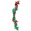
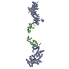
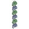
- PDB-6sl7: The Delta Calcium mutant of ALPHA-ACTININ FROM ENTAMOEBA HISTOLYTICA -
+ Open data

- Basic information
| Entry | Database: PDB / ID: 6sl7 | |||||||||
|---|---|---|---|---|---|---|---|---|---|---|
| Title | The Delta Calcium mutant of ALPHA-ACTININ FROM ENTAMOEBA HISTOLYTICA | |||||||||
 Components Components | Calponin homology domain protein putative | |||||||||
 Keywords Keywords | STRUCTURAL PROTEIN / Actinin / Actin Binding Domain / Spectrin Repear / Calmodulin-like domain / Calcium Regulation | |||||||||
| Function / homology |  Function and homology information Function and homology information | |||||||||
| Biological species |   Entamoeba histolytica (eukaryote) Entamoeba histolytica (eukaryote) | |||||||||
| Method |  X-RAY DIFFRACTION / SYNCHROTRON / MOLECULAR REPLACEMENT / Resolution: 3.3 Å X-RAY DIFFRACTION / SYNCHROTRON / MOLECULAR REPLACEMENT / Resolution: 3.3 Å | |||||||||
 Authors Authors | Pinotsis, N. / Lopez Arolas, A. / Djinovic-Carugo, K. | |||||||||
| Funding support |  Austria, 2items Austria, 2items
| |||||||||
 Citation Citation | Journal: Proc.Natl.Acad.Sci.USA / Year: 2020 Title: Calcium modulates the domain flexibility and function of an alpha-actinin similar to the ancestral alpha-actinin. Authors: Pinotsis, N. / Zielinska, K. / Babuta, M. / Arolas, J.L. / Kostan, J. / Khan, M.B. / Schreiner, C. / Salmazo, A. / Ciccarelli, L. / Puchinger, M. / Gkougkoulia, E.A. / Ribeiro Jr., E.A. / ...Authors: Pinotsis, N. / Zielinska, K. / Babuta, M. / Arolas, J.L. / Kostan, J. / Khan, M.B. / Schreiner, C. / Salmazo, A. / Ciccarelli, L. / Puchinger, M. / Gkougkoulia, E.A. / Ribeiro Jr., E.A. / Marlovits, T.C. / Bhattacharya, A. / Djinovic-Carugo, K. | |||||||||
| History |
|
- Structure visualization
| Structure viewer | Molecule: MolmilJmol/JSmol |
|---|
- Downloads & links
-Download
| PDBx/mmCIF format | 6sl7.cif.gz | 136 KB | Display | PDBx/mmCIF format |
|---|---|---|---|---|
| PDB format | pdb6sl7.ent.gz | 103.5 KB | Display | PDB format |
| PDBx/mmJSON format | 6sl7.json.gz | Tree view | PDBx/mmJSON format | |
| Others |  Other downloads Other downloads |
-Validation report
| Summary document | 6sl7_validation.pdf.gz | 431.6 KB | Display | wwPDB validaton report |
|---|---|---|---|---|
| Full document | 6sl7_full_validation.pdf.gz | 460.7 KB | Display | |
| Data in XML | 6sl7_validation.xml.gz | 25.7 KB | Display | |
| Data in CIF | 6sl7_validation.cif.gz | 34 KB | Display | |
| Arichive directory | https://data.pdbj.org/pub/pdb/validation_reports/sl/6sl7ftp://data.pdbj.org/pub/pdb/validation_reports/sl/6sl7 | HTTPS FTP |
-Related structure data
| Related structure data | 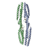 5nl6C  5nl7C  6sl2SC  6sl3C C: citing same article ( S: Starting model for refinement |
|---|---|
| Similar structure data |


-Links
 PDBj
PDBj- Assembly
| Deposited unit | 
| ||||||||||||
|---|---|---|---|---|---|---|---|---|---|---|---|---|---|
| 1 | 
| ||||||||||||
| Unit cell |
|
-Components
| #1: Protein | Mass: 69853.633 Da / Num. of mol.: 1 Source method: isolated from a genetically manipulated source Source: (gene. exp.) Entamoeba histolytica (eukaryote) / Gene: CL6EHI_199000, EHI_199000 / Production host:  |
|---|---|
| Has ligand of interest | N |
-Experimental details
-Experiment
| Experiment | Method: X-RAY DIFFRACTION / Number of used crystals: 1 |
|---|
- Sample preparation
| Crystal | Density Matthews: 2.59 Å3/Da / Density % sol: 52.49 % |
|---|---|
| Crystal grow | Temperature: 293 K / Method: vapor diffusion, hanging drop / pH: 5.5 Details: 0.1 M bis-tris (pH 5.5), 0.3 M MgCl2, 20% (w/v) PEG 3,350 |
-Data collection
| Diffraction | Mean temperature: 100 K / Serial crystal experiment: N |
|---|---|
| Diffraction source | Source: SYNCHROTRON / Site: ESRF  / Beamline: ID23-1 / Wavelength: 0.96862 Å / Beamline: ID23-1 / Wavelength: 0.96862 Å |
| Detector | Type: DECTRIS PILATUS3 S 6M / Detector: PIXEL / Date: Aug 1, 2014 |
| Radiation | Protocol: SINGLE WAVELENGTH / Monochromatic (M) / Laue (L): M / Scattering type: x-ray |
| Radiation wavelength | Wavelength: 0.96862 Å / Relative weight: 1 |
| Reflection | Resolution: 3.3→46.85 Å / Num. obs: 11494 / % possible obs: 97.8 % / Redundancy: 4.6 % / CC1/2: 0.998 / Rrim(I) all: 0.084 / Net I/σ(I): 11.57 |
| Reflection shell | Resolution: 3.3→3.38 Å / Redundancy: 5.8 % / Num. unique obs: 2235 / CC1/2: 0.855 / % possible all: 96.2 |
- Processing
| Software |
| ||||||||||||||||
|---|---|---|---|---|---|---|---|---|---|---|---|---|---|---|---|---|---|
| Refinement | Method to determine structure: MOLECULAR REPLACEMENT Starting model: 6SL2 Resolution: 3.3→46.32 Å / Cross valid method: FREE R-VALUE
| ||||||||||||||||
| Refinement step | Cycle: LAST / Resolution: 3.3→46.32 Å
|
