 Movie
Movie Controller


+ Open data

- Basic information
| Entry | Database: PDB / ID: 5v6t | |||||||||
|---|---|---|---|---|---|---|---|---|---|---|
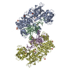
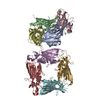
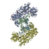
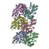
| Title | The Plexin D1 intracellular region in complex with GIPC1 | |||||||||
 Components Components |
| |||||||||
 Keywords Keywords | PROTEIN BINDING | |||||||||
| Function / homology |  Function and homology information Function and homology informationTGFBR3 regulates FGF2 signaling / FGFR1b ligand binding and activation / FGFR1c ligand binding and activation / Other semaphorin interactions / TGFBR3 regulates TGF-beta signaling / dichotomous subdivision of terminal units involved in salivary gland branching / RND2 GTPase cycle / cardiac septum development / positive regulation of melanin biosynthetic process / synaptic target recognition ...TGFBR3 regulates FGF2 signaling / FGFR1b ligand binding and activation / FGFR1c ligand binding and activation / Other semaphorin interactions / TGFBR3 regulates TGF-beta signaling / dichotomous subdivision of terminal units involved in salivary gland branching / RND2 GTPase cycle / cardiac septum development / positive regulation of melanin biosynthetic process / synaptic target recognition / semaphorin receptor complex / semaphorin receptor activity / negative regulation of cell adhesion / coronary vasculature development / glutamate secretion / vesicle membrane / cellular response to interleukin-7 / positive regulation of axonogenesis / aorta development / branching involved in blood vessel morphogenesis / myosin binding / positive regulation of cytokinesis / positive regulation of transforming growth factor beta receptor signaling pathway / outflow tract morphogenesis / semaphorin-plexin signaling pathway / lamellipodium membrane / brush border / regulation of angiogenesis / endocytic vesicle / endothelial cell migration / protein targeting / synapse assembly / negative regulation of proteasomal ubiquitin-dependent protein catabolic process / regulation of cell migration / presynaptic modulation of chemical synaptic transmission / GTPase activator activity / dendritic shaft / PDZ domain binding / kidney development / regulation of protein stability / regulation of synaptic plasticity / Schaffer collateral - CA1 synapse / synaptic vesicle / regulation of cell shape / lamellipodium / presynapse / actin binding / cell body / angiogenesis / cytoplasmic vesicle / cell cortex / dendritic spine / chemical synaptic transmission / negative regulation of neuron apoptotic process / postsynapse / G protein-coupled receptor signaling pathway / signaling receptor binding / protein domain specific binding / axon / glutamatergic synapse / membrane / identical protein binding / plasma membrane / cytoplasm / cytosol Similarity search - Function | |||||||||
| Biological species |  | |||||||||
| Method |  X-RAY DIFFRACTION / SYNCHROTRON / MOLECULAR REPLACEMENT / Resolution: 3.189 Å X-RAY DIFFRACTION / SYNCHROTRON / MOLECULAR REPLACEMENT / Resolution: 3.189 Å | |||||||||
 Authors Authors | Shang, G. / Zhang, X. | |||||||||
| Funding support |  United States, 2items United States, 2items
| |||||||||
 Citation Citation | Journal: Elife / Year: 2017 Title: Structure analyses reveal a regulated oligomerization mechanism of the PlexinD1/GIPC/myosin VI complex. Authors: Shang, G. / Brautigam, C.A. / Chen, R. / Lu, D. / Torres-Vazquez, J. / Zhang, X. | |||||||||
| History |
|
- Structure visualization
| Structure viewer | Molecule: MolmilJmol/JSmol |
|---|
- Downloads & links
-Download
| PDBx/mmCIF format | 5v6t.cif.gz | 290.2 KB | Display | PDBx/mmCIF format |
|---|---|---|---|---|
| PDB format | pdb5v6t.ent.gz | 232.2 KB | Display | PDB format |
| PDBx/mmJSON format | 5v6t.json.gz | Tree view | PDBx/mmJSON format | |
| Others |  Other downloads Other downloads |
-Validation report
| Arichive directory | https://data.pdbj.org/pub/pdb/validation_reports/v6/5v6tftp://data.pdbj.org/pub/pdb/validation_reports/v6/5v6t | HTTPS FTP |
|---|
-Related structure data
| Related structure data | 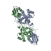 5v6bC 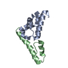 5v6eC 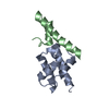 5v6hC 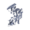 5v6rSC C: citing same article ( S: Starting model for refinement |
|---|---|
| Similar structure data |


-Links
 PDBj
PDBj


- Assembly
| Deposited unit | 
| ||||||||
|---|---|---|---|---|---|---|---|---|---|
| 1 | 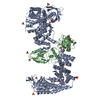
| ||||||||
| 2 | 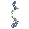
| ||||||||
| Unit cell |
|
-Components
| #1: Protein | Mass: 67870.641 Da / Num. of mol.: 1 / Fragment: UNP residues 1339-1925 Source method: isolated from a genetically manipulated source Source: (gene. exp.)  | ||
|---|---|---|---|
| #2: Protein | Mass: 31541.020 Da / Num. of mol.: 1 / Fragment: UNP residues 52-333 Source method: isolated from a genetically manipulated source Source: (gene. exp.) | ||
| #3: Chemical | ChemComp-SO4 /   Mass: 96.063 Da / Num. of mol.: 7 / Source method: obtained synthetically / Formula: SO4 Mass: 96.063 Da / Num. of mol.: 7 / Source method: obtained synthetically / Formula: SO4#4: Water | ChemComp-HOH / |  Mass: 18.015 Da / Num. of mol.: 19 / Source method: isolated from a natural source / Formula: H2O Mass: 18.015 Da / Num. of mol.: 19 / Source method: isolated from a natural source / Formula: H2O |
-Experimental details
-Experiment
| Experiment | Method: X-RAY DIFFRACTION / Number of used crystals: 1 |
|---|
- Sample preparation
| Crystal | Density Matthews: 3.84 Å3/Da / Density % sol: 68.01 % |
|---|---|
| Crystal grow | Temperature: 293 K / Method: vapor diffusion, sitting drop / Details: 0.1 M Bis-Tris pH 5.5 and 2 M (NH4)2SO4 |
-Data collection
| Diffraction | Mean temperature: 100 K |
|---|---|
| Diffraction source | Source: SYNCHROTRON / Site: APS / Beamline: 19-ID / Wavelength: 0.9794 Å |
| Detector | Type: ADSC QUANTUM 315 / Detector: CCD / Date: Jun 15, 2015 |
| Radiation | Protocol: SINGLE WAVELENGTH / Monochromatic (M) / Laue (L): M / Scattering type: x-ray |
| Radiation wavelength | Wavelength: 0.9794 Å / Relative weight: 1 |
| Reflection | Resolution: 3.2→50 Å / Num. obs: 27629 / % possible obs: 100 % / Redundancy: 12 % / Rpim(I) all: 0.041 / Net I/σ(I): 17.5 |
| Reflection shell | Resolution: 3.2→3.26 Å / Redundancy: 5.6 % / Mean I/σ(I) obs: 2 / Rpim(I) all: 0.361 / % possible all: 100 |
- Processing
| Software |
| ||||||||||||||||||||||||||||||||||||||||||||||||||||||||||||||||||||||||||||||||||||||||||||||||||
|---|---|---|---|---|---|---|---|---|---|---|---|---|---|---|---|---|---|---|---|---|---|---|---|---|---|---|---|---|---|---|---|---|---|---|---|---|---|---|---|---|---|---|---|---|---|---|---|---|---|---|---|---|---|---|---|---|---|---|---|---|---|---|---|---|---|---|---|---|---|---|---|---|---|---|---|---|---|---|---|---|---|---|---|---|---|---|---|---|---|---|---|---|---|---|---|---|---|---|---|
| Refinement | Method to determine structure: MOLECULAR REPLACEMENT Starting model: 5V6R Resolution: 3.189→49.033 Å / SU ML: 0.34 / Cross valid method: FREE R-VALUE / σ(F): 1.5 / Phase error: 19.96 / Stereochemistry target values: ML
| ||||||||||||||||||||||||||||||||||||||||||||||||||||||||||||||||||||||||||||||||||||||||||||||||||
| Solvent computation | Shrinkage radii: 0.9 Å / VDW probe radii: 1.11 Å / Solvent model: FLAT BULK SOLVENT MODEL | ||||||||||||||||||||||||||||||||||||||||||||||||||||||||||||||||||||||||||||||||||||||||||||||||||
| Refinement step | Cycle: LAST / Resolution: 3.189→49.033 Å
| ||||||||||||||||||||||||||||||||||||||||||||||||||||||||||||||||||||||||||||||||||||||||||||||||||
| Refine LS restraints |
| ||||||||||||||||||||||||||||||||||||||||||||||||||||||||||||||||||||||||||||||||||||||||||||||||||
| LS refinement shell |
| ||||||||||||||||||||||||||||||||||||||||||||||||||||||||||||||||||||||||||||||||||||||||||||||||||
| Refinement TLS params. | Method: refined / Refine-ID: X-RAY DIFFRACTION
| ||||||||||||||||||||||||||||||||||||||||||||||||||||||||||||||||||||||||||||||||||||||||||||||||||
| Refinement TLS group |
|
