 Movie
Movie Controller

[English] 日本語
 Yorodumi
- PDB-6sav: Structural and functional characterisation of three novel fungal ... -
+ Open data

- Basic information
| Entry | Database: PDB / ID: 6sav | ||||||
|---|---|---|---|---|---|---|---|
| Title | Structural and functional characterisation of three novel fungal amylases with enhanced stability and pH tolerance | ||||||
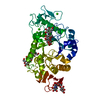
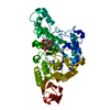
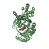
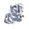
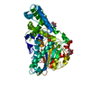
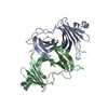
 Components Components | (Alpha-amylase) x 2 | ||||||
 Keywords Keywords | HYDROLASE / biotechnology / fungal amylase | ||||||
| Function / homology |  Function and homology information Function and homology informationalpha-amylase / alpha-amylase activity / carbohydrate catabolic process / calcium ion binding Similarity search - Function | ||||||
| Biological species |  Rhizomucor pusillus (fungus) Rhizomucor pusillus (fungus) | ||||||
| Method |  X-RAY DIFFRACTION / SYNCHROTRON / MOLECULAR REPLACEMENT / Resolution: 1.4 Å X-RAY DIFFRACTION / SYNCHROTRON / MOLECULAR REPLACEMENT / Resolution: 1.4 Å | ||||||
 Authors Authors | Roth, C. / Moroz, O.V. / Turkenburg, J.P. / Blagova, E. / Waterman, J. / Ariza, A. / Ming, L. / Tianqi, S. / Andersen, C. / Davies, G.J. / Wilson, K.S. | ||||||
 Citation Citation | Journal: Int J Mol Sci / Year: 2019 Title: Structural and Functional Characterization of Three Novel Fungal Amylases with Enhanced Stability and pH Tolerance. Authors: Roth, C. / Moroz, O.V. / Turkenburg, J.P. / Blagova, E. / Waterman, J. / Ariza, A. / Ming, L. / Tianqi, S. / Andersen, C. / Davies, G.J. / Wilson, K.S. | ||||||
| History |
|
- Structure visualization
| Structure viewer | Molecule: MolmilJmol/JSmol |
|---|
- Downloads & links
-Download
| PDBx/mmCIF format | 6sav.cif.gz | 217.9 KB | Display | PDBx/mmCIF format |
|---|---|---|---|---|
| PDB format | pdb6sav.ent.gz | 169.2 KB | Display | PDB format |
| PDBx/mmJSON format | 6sav.json.gz | Tree view | PDBx/mmJSON format | |
| Others |  Other downloads Other downloads |
-Validation report
| Arichive directory | https://data.pdbj.org/pub/pdb/validation_reports/sa/6savftp://data.pdbj.org/pub/pdb/validation_reports/sa/6sav | HTTPS FTP |
|---|
-Related structure data
| Related structure data | 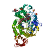 6saoC 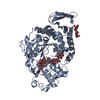 6sauC 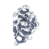 2taaS S: Starting model for refinement C: citing same article ( |
|---|---|
| Similar structure data |


-Links
 PDBj
PDBj

- Assembly
| Deposited unit | 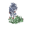
| ||||||||
|---|---|---|---|---|---|---|---|---|---|
| 1 | 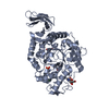
| ||||||||
| 2 | 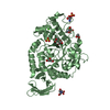
| ||||||||
| Unit cell |
|
-Components
-Protein , 2 types, 2 molecules AB
| #1: Protein | Mass: 48268.855 Da / Num. of mol.: 1 Source method: isolated from a genetically manipulated source Source: (gene. exp.) Rhizomucor pusillus (fungus) / Production host: |
|---|---|
| #2: Protein | Mass: 48269.840 Da / Num. of mol.: 1 Source method: isolated from a genetically manipulated source Source: (gene. exp.) Rhizomucor pusillus (fungus) / Production host: |
-Sugars , 1 types, 3 molecules 
| #4: Sugar |  Type: D-saccharide, beta linking / Mass: 221.208 Da / Num. of mol.: 3 Type: D-saccharide, beta linking / Mass: 221.208 Da / Num. of mol.: 3Source method: isolated from a genetically manipulated source Formula: C8H15NO6 |
|---|
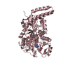
-Non-polymers , 4 types, 955 molecules 


| #3: Chemical |  Mass: 40.078 Da / Num. of mol.: 2 / Source method: obtained synthetically / Formula: Ca Mass: 40.078 Da / Num. of mol.: 2 / Source method: obtained synthetically / Formula: Ca#5: Chemical | ChemComp-GOL /  Mass: 92.094 Da / Num. of mol.: 7 / Source method: obtained synthetically / Formula: C3H8O3 Mass: 92.094 Da / Num. of mol.: 7 / Source method: obtained synthetically / Formula: C3H8O3#6: Chemical |  Mass: 96.063 Da / Num. of mol.: 3 / Source method: obtained synthetically / Formula: SO4 Mass: 96.063 Da / Num. of mol.: 3 / Source method: obtained synthetically / Formula: SO4#7: Water | ChemComp-HOH / | Mass: 18.015 Da / Num. of mol.: 943 / Source method: isolated from a natural source / Formula: H2O |
|---|
-Details
| Has ligand of interest | N |
|---|---|
| Has protein modification | Y |
-Experimental details
-Experiment
| Experiment | Method: X-RAY DIFFRACTION / Number of used crystals: 1 |
|---|
- Sample preparation
| Crystal | Density Matthews: 2.13 Å3/Da / Density % sol: 42.39 % |
|---|---|
| Crystal grow | Temperature: 292 K / Method: vapor diffusion, hanging drop / pH: 4.6 / Details: 0.2 M NaCl, 0.1 M Na-acetate pH 4.6, 30 %MPD |
-Data collection
| Diffraction | Mean temperature: 100 K / Serial crystal experiment: N | |||||||||||||||||||||||||||
|---|---|---|---|---|---|---|---|---|---|---|---|---|---|---|---|---|---|---|---|---|---|---|---|---|---|---|---|---|
| Diffraction source | Source: SYNCHROTRON / Site: ESRF  / Beamline: ID29 / Wavelength: 1.0004 Å / Beamline: ID29 / Wavelength: 1.0004 Å | |||||||||||||||||||||||||||
| Detector | Type: MARRESEARCH / Detector: CCD / Date: Mar 17, 2007 | |||||||||||||||||||||||||||
| Radiation | Protocol: SINGLE WAVELENGTH / Monochromatic (M) / Laue (L): M / Scattering type: x-ray | |||||||||||||||||||||||||||
| Radiation wavelength | Wavelength: 1.0004 Å / Relative weight: 1 | |||||||||||||||||||||||||||
| Reflection | Resolution: 1.4→43.21 Å / Num. obs: 146177 / % possible obs: 92.9 % / Redundancy: 2.2 % / CC1/2: 0.998 / Rmerge(I) obs: 0.033 / Rpim(I) all: 0.03 / Rrim(I) all: 0.044 / Net I/σ(I): 9.6 / Num. measured all: 315876 / Scaling rejects: 5 | |||||||||||||||||||||||||||
| Reflection shell | Diffraction-ID: 1 / Redundancy: 2.1 %
|
- Processing
| Software |
| ||||||||||||||||||||||||||||||||||||||||||||||||||||||||||||
|---|---|---|---|---|---|---|---|---|---|---|---|---|---|---|---|---|---|---|---|---|---|---|---|---|---|---|---|---|---|---|---|---|---|---|---|---|---|---|---|---|---|---|---|---|---|---|---|---|---|---|---|---|---|---|---|---|---|---|---|---|---|
| Refinement | Method to determine structure: MOLECULAR REPLACEMENT Starting model: 2taa Resolution: 1.4→39.99 Å / Cor.coef. Fo:Fc: 0.976 / Cor.coef. Fo:Fc free: 0.964 / SU B: 0.92 / SU ML: 0.036 / Cross valid method: THROUGHOUT / σ(F): 0 / ESU R: 0.056 / ESU R Free: 0.059 / Stereochemistry target values: MAXIMUM LIKELIHOOD Details: HYDROGENS HAVE BEEN ADDED IN THE RIDING POSITIONS U VALUES : REFINED INDIVIDUALLY
| ||||||||||||||||||||||||||||||||||||||||||||||||||||||||||||
| Solvent computation | Ion probe radii: 0.8 Å / Shrinkage radii: 0.8 Å / VDW probe radii: 1.2 Å / Solvent model: MASK | ||||||||||||||||||||||||||||||||||||||||||||||||||||||||||||
| Displacement parameters | Biso max: 74.22 Å2 / Biso mean: 12.475 Å2 / Biso min: 5.91 Å2
| ||||||||||||||||||||||||||||||||||||||||||||||||||||||||||||
| Refinement step | Cycle: final / Resolution: 1.4→39.99 Å
| ||||||||||||||||||||||||||||||||||||||||||||||||||||||||||||
| Refine LS restraints |
| ||||||||||||||||||||||||||||||||||||||||||||||||||||||||||||
| LS refinement shell | Resolution: 1.4→1.436 Å / Rfactor Rfree error: 0 / Total num. of bins used: 20
|
