 Movie
Movie Controller

[English] 日本語
 Yorodumi
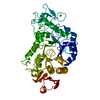
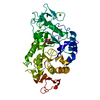
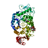
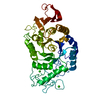
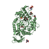
- PDB-1rp9: Crystal structure of barley alpha-amylase isozyme 1 (amy1) inacti... -
+ Open data

- Basic information
| Entry | Database: PDB / ID: 1rp9 | |||||||||
|---|---|---|---|---|---|---|---|---|---|---|
| Title | Crystal structure of barley alpha-amylase isozyme 1 (amy1) inactive mutant d180a in complex with acarbose | |||||||||
 Components Components | Alpha-amylase type 1 isozyme | |||||||||
 Keywords Keywords | HYDROLASE / ALPHA-AMYLASE / BARLEY / ISOZYME 1 / INACTIVE MUTANT / BETA-ALPHA-BARREL / SUGAR TONGS BINDING SITE / ACARBOSE | |||||||||
| Function / homology |  Function and homology information Function and homology informationstarch catabolic process / alpha-amylase / alpha-amylase activity / calcium ion binding / extracellular region Similarity search - Function | |||||||||
| Biological species |  | |||||||||
| Method |  X-RAY DIFFRACTION / SYNCHROTRON / FOURIER DIFFERENCE / Resolution: 2 Å X-RAY DIFFRACTION / SYNCHROTRON / FOURIER DIFFERENCE / Resolution: 2 Å | |||||||||
 Authors Authors | Robert, X. / Haser, R. / Aghajari, N. | |||||||||
 Citation Citation | Journal: J.Biol.Chem. / Year: 2005 Title: Oligosaccharide Binding to Barley {alpha}-Amylase 1 Authors: Robert, X. / Haser, R. / Mori, H. / Svensson, B. / Aghajari, N. | |||||||||
| History |
| |||||||||
| Remark 999 | SEQUENCE ACCORDING TO THE AUTHORS, THERE IS AN ERROR IN SWISSPROT DATABASE, CORRECTED BY ...SEQUENCE ACCORDING TO THE AUTHORS, THERE IS AN ERROR IN SWISSPROT DATABASE, CORRECTED BY EXAMINATION OF THE CRYSTAL STRUCTURE. |
- Structure visualization
| Structure viewer | Molecule: MolmilJmol/JSmol |
|---|
- Downloads & links
-Download
| PDBx/mmCIF format | 1rp9.cif.gz | 105.8 KB | Display | PDBx/mmCIF format |
|---|---|---|---|---|
| PDB format | pdb1rp9.ent.gz | 78.6 KB | Display | PDB format |
| PDBx/mmJSON format | 1rp9.json.gz | Tree view | PDBx/mmJSON format | |
| Others |  Other downloads Other downloads |
-Validation report
| Arichive directory | https://data.pdbj.org/pub/pdb/validation_reports/rp/1rp9ftp://data.pdbj.org/pub/pdb/validation_reports/rp/1rp9 | HTTPS FTP |
|---|
-Related structure data
| Related structure data | 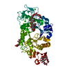 1rp8C  1rpkC  1ht6S S: Starting model for refinement C: citing same article ( |
|---|---|
| Similar structure data |


-Links
 PDBj
PDBj

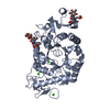
- Assembly
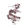
| Deposited unit | 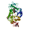
| ||||||||
|---|---|---|---|---|---|---|---|---|---|
| 1 |
| ||||||||
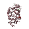
| Unit cell |
|
-Components
| #1: Protein | Mass: 44596.047 Da / Num. of mol.: 1 / Mutation: D180A Source method: isolated from a genetically manipulated source Source: (gene. exp.)  Pichia pastoris (fungus) / Strain (production host): GS115 / References: UniProt: P00693, alpha-amylase Pichia pastoris (fungus) / Strain (production host): GS115 / References: UniProt: P00693, alpha-amylase | ||||
|---|---|---|---|---|---|
| #2: Polysaccharide | 4,6-dideoxy-4-{[(1S,5R,6S)-3-formyl-5,6-dihydroxy-4-oxocyclohex-2-en-1-yl]amino}-alpha-D-xylo-hex-5- ...4,6-dideoxy-4-{[(1S,5R,6S)-3-formyl-5,6-dihydroxy-4-oxocyclohex-2-en-1-yl]amino}-alpha-D-xylo-hex-5-enopyranose-(1-4)-beta-D-glucopyranose-(1-4)-alpha-D-glucopyranose Type: oligosaccharide / Mass: 639.558 Da / Num. of mol.: 1 Source method: isolated from a genetically manipulated source | ||||
| #3: Polysaccharide | Type: oligosaccharide / Mass: 639.558 Da / Num. of mol.: 2 Source method: isolated from a genetically manipulated source #4: Chemical |   Mass: 40.078 Da / Num. of mol.: 3 / Source method: obtained synthetically / Formula: Ca Mass: 40.078 Da / Num. of mol.: 3 / Source method: obtained synthetically / Formula: Ca#5: Water | ChemComp-HOH / |  Mass: 18.015 Da / Num. of mol.: 396 / Source method: isolated from a natural source / Formula: H2O Mass: 18.015 Da / Num. of mol.: 396 / Source method: isolated from a natural source / Formula: H2O |
-Experimental details
-Experiment
| Experiment | Method: X-RAY DIFFRACTION / Number of used crystals: 1 |
|---|
- Sample preparation
| Crystal | Density Matthews: 2.36 Å3/Da / Density % sol: 47.85 % |
|---|---|
| Crystal grow | Temperature: 290 K / Method: vapor diffusion, hanging drop / pH: 6.7 Details: PEG 8000, ISOPROPANOL, pH 6.7, VAPOR DIFFUSION, HANGING DROP, temperature 290K |
-Data collection
| Diffraction | Mean temperature: 100 K |
|---|---|
| Diffraction source | Source: SYNCHROTRON / Site: ESRF  / Beamline: BM30A / Wavelength: 0.95 Å / Beamline: BM30A / Wavelength: 0.95 Å |
| Detector | Type: MARRESEARCH / Detector: CCD / Date: Dec 13, 2000 |
| Radiation | Monochromator: MIRRORS / Protocol: SINGLE WAVELENGTH / Monochromatic (M) / Laue (L): M / Scattering type: x-ray |
| Radiation wavelength | Wavelength: 0.95 Å / Relative weight: 1 |
| Reflection | Resolution: 2→39.52 Å / Num. all: 25774 / Num. obs: 25774 / % possible obs: 100 % / Observed criterion σ(F): 0 / Observed criterion σ(I): 0 / Redundancy: 4.4 % / Biso Wilson estimate: 13 Å2 / Rsym value: 0.083 / Net I/σ(I): 7.5 |
| Reflection shell | Resolution: 2→2.05 Å / Redundancy: 4.4 % / Mean I/σ(I) obs: 2.4 / Num. unique all: 1891 / Rsym value: 0.315 / % possible all: 100 |
- Processing
| Software |
| ||||||||||||||||||||||||||||||||||||
|---|---|---|---|---|---|---|---|---|---|---|---|---|---|---|---|---|---|---|---|---|---|---|---|---|---|---|---|---|---|---|---|---|---|---|---|---|---|
| Refinement | Method to determine structure: FOURIER DIFFERENCE Starting model: PDB ENTRY 1HT6 Resolution: 2→39.52 Å / Data cutoff high absF: 1631246.97 / Data cutoff low absF: 0 / Isotropic thermal model: RESTRAINED / Cross valid method: THROUGHOUT / σ(F): 0 / σ(I): 0 / Stereochemistry target values: Engh & Huber
| ||||||||||||||||||||||||||||||||||||
| Displacement parameters | Biso mean: 21.4 Å2
| ||||||||||||||||||||||||||||||||||||
| Refine analyze |
| ||||||||||||||||||||||||||||||||||||
| Refinement step | Cycle: LAST / Resolution: 2→39.52 Å
| ||||||||||||||||||||||||||||||||||||
| Refine LS restraints |
| ||||||||||||||||||||||||||||||||||||
| LS refinement shell | Resolution: 2→2.13 Å / Rfactor Rfree error: 0.011 / Total num. of bins used: 6
| ||||||||||||||||||||||||||||||||||||
| Xplor file |
|
