 Movie
Movie Controller

[English] 日本語
 Yorodumi
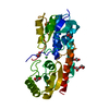
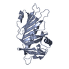
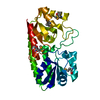
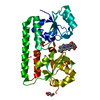
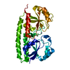
- PDB-6ru4: Structure of the SBP FpvC from pseudomonas aeruginosa in complex ... -
+ Open data

- Basic information
| Entry | Database: PDB / ID: 6ru4 | ||||||
|---|---|---|---|---|---|---|---|
| Title | Structure of the SBP FpvC from pseudomonas aeruginosa in complex with Mn2+ | ||||||
 Components Components | Probable adhesion protein | ||||||
 Keywords Keywords | TRANSPORT PROTEIN | ||||||
| Function / homology |  Function and homology information Function and homology information | ||||||
| Biological species |  Pseudomonas aeruginosa PAO1 (bacteria) Pseudomonas aeruginosa PAO1 (bacteria) | ||||||
| Method |  X-RAY DIFFRACTION / SYNCHROTRON / MOLECULAR REPLACEMENT / Resolution: 2.49 Å X-RAY DIFFRACTION / SYNCHROTRON / MOLECULAR REPLACEMENT / Resolution: 2.49 Å | ||||||
 Authors Authors | Morera, S. / Vigouroux, A. | ||||||
 Citation Citation | Journal: Febs J. / Year: 2020 Title: A unique ferrous iron binding mode is associated with large conformational changes for the transport protein FpvC of Pseudomonas aeruginosa. Authors: Vigouroux, A. / Aumont-Nicaise, M. / Boussac, A. / Marty, L. / Lo Bello, L. / Legrand, P. / Brillet, K. / Schalk, I.J. / Morera, S. | ||||||
| History |
|
- Structure visualization
| Structure viewer | Molecule: MolmilJmol/JSmol |
|---|
- Downloads & links
-Download
| PDBx/mmCIF format | 6ru4.cif.gz | 431.2 KB | Display | PDBx/mmCIF format |
|---|---|---|---|---|
| PDB format | pdb6ru4.ent.gz | 356.8 KB | Display | PDB format |
| PDBx/mmJSON format | 6ru4.json.gz | Tree view | PDBx/mmJSON format | |
| Others |  Other downloads Other downloads |
-Validation report
| Summary document | 6ru4_validation.pdf.gz | 7.8 MB | Display | wwPDB validaton report |
|---|---|---|---|---|
| Full document | 6ru4_full_validation.pdf.gz | 7.8 MB | Display | |
| Data in XML | 6ru4_validation.xml.gz | 40 KB | Display | |
| Data in CIF | 6ru4_validation.cif.gz | 56.4 KB | Display | |
| Arichive directory | https://data.pdbj.org/pub/pdb/validation_reports/ru/6ru4ftp://data.pdbj.org/pub/pdb/validation_reports/ru/6ru4 | HTTPS FTP |
-Related structure data
| Related structure data |  6r3zSC  6r44C  6r5sC  6r6kC S: Starting model for refinement C: citing same article ( |
|---|---|
| Similar structure data |


-Links
 PDBj
PDBj- Assembly
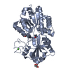
| Deposited unit | 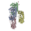
| ||||||||
|---|---|---|---|---|---|---|---|---|---|
| 1 | 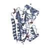
| ||||||||
| 2 | 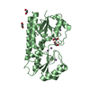
| ||||||||
| 3 | 
| ||||||||
| 4 | 
| ||||||||
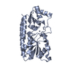
| Unit cell |
|
-Components
| #1: Protein | Mass: 30629.920 Da / Num. of mol.: 4 Source method: isolated from a genetically manipulated source Source: (gene. exp.) Pseudomonas aeruginosa PAO1 (bacteria) / Gene: PA2407Variant: ATCC 15692 / DSM 22644 / CIP 104116 / JCM 14847 / LMG 12228 / 1C / PRS 101 / PAO1 Production host: #2: Chemical | ChemComp-MN /   Mass: 54.938 Da / Num. of mol.: 28 / Source method: obtained synthetically / Formula: Mn / Feature type: SUBJECT OF INVESTIGATION Mass: 54.938 Da / Num. of mol.: 28 / Source method: obtained synthetically / Formula: Mn / Feature type: SUBJECT OF INVESTIGATION#3: Chemical | ChemComp-EDO /   Mass: 62.068 Da / Num. of mol.: 7 / Source method: obtained synthetically / Formula: C2H6O2 / Feature type: SUBJECT OF INVESTIGATION Mass: 62.068 Da / Num. of mol.: 7 / Source method: obtained synthetically / Formula: C2H6O2 / Feature type: SUBJECT OF INVESTIGATION#4: Chemical |   Mass: 106.120 Da / Num. of mol.: 2 / Source method: obtained synthetically / Formula: C4H10O3 / Feature type: SUBJECT OF INVESTIGATION Mass: 106.120 Da / Num. of mol.: 2 / Source method: obtained synthetically / Formula: C4H10O3 / Feature type: SUBJECT OF INVESTIGATION#5: Water | ChemComp-HOH / |  Mass: 18.015 Da / Num. of mol.: 199 / Source method: isolated from a natural source / Formula: H2O Mass: 18.015 Da / Num. of mol.: 199 / Source method: isolated from a natural source / Formula: H2O |
|---|
-Experimental details
-Experiment
| Experiment | Method: X-RAY DIFFRACTION / Number of used crystals: 1 |
|---|
- Sample preparation
| Crystal | Density Matthews: 2.14 Å3/Da / Density % sol: 42.51 % |
|---|---|
| Crystal grow | Temperature: 292 K / Method: vapor diffusion, hanging drop / pH: 6.5 / Details: MPEG 2K, 0.1 M MES |
-Data collection
| Diffraction | Mean temperature: 100 K / Serial crystal experiment: N |
|---|---|
| Diffraction source | Source: SYNCHROTRON / Site: SOLEIL  / Beamline: PROXIMA 2 / Wavelength: 0.98 / Wavelength: 0.98 Å / Beamline: PROXIMA 2 / Wavelength: 0.98 / Wavelength: 0.98 Å |
| Detector | Type: DECTRIS EIGER X 16M / Detector: PIXEL / Date: Apr 19, 2019 |
| Radiation | Protocol: SINGLE WAVELENGTH / Monochromatic (M) / Laue (L): M / Scattering type: x-ray |
| Radiation wavelength | Wavelength: 0.98 Å / Relative weight: 1 |
| Reflection | Resolution: 2.49→44.57 Å / Num. obs: 35191 / % possible obs: 98.4 % / Redundancy: 6.9 % / Biso Wilson estimate: 71.47 Å2 / CC1/2: 0.996 / Rpim(I) all: 0.07 / Rsym value: 0.17 / Net I/σ(I): 8.58 |
| Reflection shell | Resolution: 2.49→2.64 Å / Num. unique obs: 5188 |
- Processing
| Software |
| |||||||||||||||||||||||||||||||||||||||||||||||||||||||||||||||||||||||||||||||||||||||||||||||||||||||||||||||||||||||||||||
|---|---|---|---|---|---|---|---|---|---|---|---|---|---|---|---|---|---|---|---|---|---|---|---|---|---|---|---|---|---|---|---|---|---|---|---|---|---|---|---|---|---|---|---|---|---|---|---|---|---|---|---|---|---|---|---|---|---|---|---|---|---|---|---|---|---|---|---|---|---|---|---|---|---|---|---|---|---|---|---|---|---|---|---|---|---|---|---|---|---|---|---|---|---|---|---|---|---|---|---|---|---|---|---|---|---|---|---|---|---|---|---|---|---|---|---|---|---|---|---|---|---|---|---|---|---|---|
| Refinement | Method to determine structure: MOLECULAR REPLACEMENT Starting model: 6R3Z Resolution: 2.49→44.57 Å / Cor.coef. Fo:Fc: 0.946 / Cor.coef. Fo:Fc free: 0.925 / SU R Cruickshank DPI: 1.689 / Cross valid method: THROUGHOUT / σ(F): 0 / SU R Blow DPI: 0.929 / SU Rfree Blow DPI: 0.283 / SU Rfree Cruickshank DPI: 0.294
| |||||||||||||||||||||||||||||||||||||||||||||||||||||||||||||||||||||||||||||||||||||||||||||||||||||||||||||||||||||||||||||
| Displacement parameters | Biso mean: 64.04 Å2
| |||||||||||||||||||||||||||||||||||||||||||||||||||||||||||||||||||||||||||||||||||||||||||||||||||||||||||||||||||||||||||||
| Refine analyze | Luzzati coordinate error obs: 0.34 Å | |||||||||||||||||||||||||||||||||||||||||||||||||||||||||||||||||||||||||||||||||||||||||||||||||||||||||||||||||||||||||||||
| Refinement step | Cycle: 1 / Resolution: 2.49→44.57 Å
| |||||||||||||||||||||||||||||||||||||||||||||||||||||||||||||||||||||||||||||||||||||||||||||||||||||||||||||||||||||||||||||
| Refine LS restraints |
| |||||||||||||||||||||||||||||||||||||||||||||||||||||||||||||||||||||||||||||||||||||||||||||||||||||||||||||||||||||||||||||
| LS refinement shell | Resolution: 2.49→2.52 Å / Total num. of bins used: 50
| |||||||||||||||||||||||||||||||||||||||||||||||||||||||||||||||||||||||||||||||||||||||||||||||||||||||||||||||||||||||||||||
| Refinement TLS params. | Method: refined / Refine-ID: X-RAY DIFFRACTION
| |||||||||||||||||||||||||||||||||||||||||||||||||||||||||||||||||||||||||||||||||||||||||||||||||||||||||||||||||||||||||||||
| Refinement TLS group |
|
