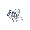
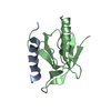
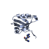
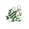
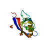
SIGNALING PROTEIN / polymeriser / plant protein / ubiquitin-like fold / polarity protein
Function / homology
Function and homology information
regulation of auxin biosynthetic process / specification of plant organ axis polarity / plant organ morphogenesis / regulation of root morphogenesis / regulation of leaf development / regulation of auxin mediated signaling pathway / inflorescence development / auxin-activated signaling pathway / response to salt / response to osmotic stress ...regulation of auxin biosynthetic process / specification of plant organ axis polarity / plant organ morphogenesis / regulation of root morphogenesis / regulation of leaf development / regulation of auxin mediated signaling pathway / inflorescence development / auxin-activated signaling pathway / response to salt / response to osmotic stress / protein polymerization / regulation of cell division / cell division / protein homodimerization activity / plasma membrane Similarity search - Function
Evidence: gel filtration, the wild type domain forms concentration dependent polymers, which can be observed in the crystal even in the polymerisation mutant (D85A)
Type
Name
Symmetry operation
Number
identity operation
1_555
x,y,z
1
Buried area
180 Å2
ΔGint
-10 kcal/mol
Surface area
5780 Å2
Method
PISA
Unit cell
Length a, b, c (Å)
47.659, 47.659, 67.971
Angle α, β, γ (deg.)
90.000, 90.000, 120.000
Int Tables number
169
Space group name H-M
P61
-
Components
#1: Protein
UPSTREAMOFFLC-likeprotein (DUF966)
Mass: 11138.453 Da / Num. of mol.: 1 / Mutation: D85A Source method: isolated from a genetically manipulated source Source: (gene. exp.) Arabidopsis thaliana (thale cress) / Gene: At3g46110 / Plasmid: pLipK Details (production host): pET based vector expressing 6xHis and Lipoyl-tag separated by TEV cleavage site Production host: Escherichia coli BL21(DE3) (bacteria) / Variant (production host): pRARE2 / References: UniProt: F4J7X6, UniProt: Q8GY65*PLUS
Method to determine structure: SAD Starting model: model Resolution: 1.7→35.28 Å / Cor.coef. Fo:Fc: 0.963 / Cor.coef. Fo:Fc free: 0.953 / SU B: 4.242 / SU ML: 0.07 / Cross valid method: THROUGHOUT / σ(F): 0 / ESU R: 0.119 / ESU R Free: 0.111 Details: HYDROGENS HAVE BEEN USED IF PRESENT IN THE INPUT U VALUES : WITH TLS ADDED
Rfactor
Num. reflection
% reflection
Selection details
Rfree
0.2145
512
5.3 %
RANDOM
Rwork
0.1846
-
-
-
obs
0.1861
9612
99.45 %
-
Solvent computation
Ion probe radii: 0.8 Å / Shrinkage radii: 0.8 Å / VDW probe radii: 1.2 Å
In the structure databanks used in Yorodumi, some data are registered as the other names, "COVID-19 virus" and "2019-nCoV". Here are the details of the virus and the list of structure data.
Jan 31, 2019. EMDB accession codes are about to change! (news from PDBe EMDB page)
EMDB accession codes are about to change! (news from PDBe EMDB page)
The allocation of 4 digits for EMDB accession codes will soon come to an end. Whilst these codes will remain in use, new EMDB accession codes will include an additional digit and will expand incrementally as the available range of codes is exhausted. The current 4-digit format prefixed with “EMD-” (i.e. EMD-XXXX) will advance to a 5-digit format (i.e. EMD-XXXXX), and so on. It is currently estimated that the 4-digit codes will be depleted around Spring 2019, at which point the 5-digit format will come into force.
The EM Navigator/Yorodumi systems omit the EMD- prefix.
Related info.:Q: What is EMD? / ID/Accession-code notation in Yorodumi/EM Navigator
Yorodumi is a browser for structure data from EMDB, PDB, SASBDB, etc.
This page is also the successor to EM Navigator detail page, and also detail information page/front-end page for Omokage search.
The word "yorodu" (or yorozu) is an old Japanese word meaning "ten thousand". "mi" (miru) is to see.
Related info.:EMDB / PDB / SASBDB / Comparison of 3 databanks / Yorodumi Search / Aug 31, 2016. New EM Navigator & Yorodumi / Yorodumi Papers / Jmol/JSmol / Function and homology information / Changes in new EM Navigator and Yorodumi
 Movie
Movie Controller


 Open data
 Basic information
 Components
 Keywords
 Function and homology information
 X-RAY DIFFRACTION /
  Authors
 United Kingdom, 1items
  Citation
 Structure visualization
 Downloads & links
 Other downloads


 PDBj
PDBj Assembly
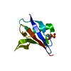


 Mass: 96.063 Da / Num. of mol.: 1 / Source method: isolated from a natural source / Formula: SO4
 Mass: 18.015 Da / Num. of mol.: 38 / Source method: isolated from a natural source / Formula: H2O
 Sample preparation
 Processing