 Movie
Movie Controller


+ Open data

- Basic information
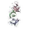
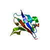
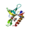
| Entry | Database: PDB / ID: 2d5g | ||||||
|---|---|---|---|---|---|---|---|
| Title | Structure of ubiquitin fold protein R767E mutant | ||||||
 Components Components | Axin-1 | ||||||
 Keywords Keywords | SIGNALING PROTEIN / UBIQUITIN FOLD | ||||||
| Function / homology |  Function and homology information Function and homology informationBeta-catenin phosphorylation cascade / Disassembly of the destruction complex and recruitment of AXIN to the membrane / TCF dependent signaling in response to WNT / Degradation of beta-catenin by the destruction complex / Degradation of AXIN / beta-catenin destruction complex assembly / armadillo repeat domain binding / head development / Ub-specific processing proteases / axial mesoderm formation ...Beta-catenin phosphorylation cascade / Disassembly of the destruction complex and recruitment of AXIN to the membrane / TCF dependent signaling in response to WNT / Degradation of beta-catenin by the destruction complex / Degradation of AXIN / beta-catenin destruction complex assembly / armadillo repeat domain binding / head development / Ub-specific processing proteases / axial mesoderm formation / cell development / dorsal/ventral axis specification / positive regulation of JUN kinase activity / response to quercetin / post-anal tail morphogenesis / beta-catenin destruction complex / dorsal/ventral pattern formation / I-SMAD binding / Wnt signalosome / axial mesoderm development / negative regulation of protein metabolic process / regulation of canonical Wnt signaling pathway / epigenetic programming in the zygotic pronuclei / nucleocytoplasmic transport / positive regulation of ubiquitin-dependent protein catabolic process / negative regulation of fat cell differentiation / positive regulation of protein kinase activity / positive regulation of transforming growth factor beta receptor signaling pathway / SMAD binding / regulation of protein phosphorylation / negative regulation of transcription elongation by RNA polymerase II / R-SMAD binding / negative regulation of Wnt signaling pathway / canonical Wnt signaling pathway / lateral plasma membrane / ubiquitin-like ligase-substrate adaptor activity / cytoplasmic microtubule organization / signaling adaptor activity / protein catabolic process / positive regulation of protein ubiquitination / protein serine/threonine kinase activator activity / protein serine/threonine kinase binding / cell periphery / sensory perception of sound / negative regulation of canonical Wnt signaling pathway / positive regulation of protein phosphorylation / positive regulation of JNK cascade / beta-catenin binding / Wnt signaling pathway / protein polyubiquitination / positive regulation of protein catabolic process / p53 binding / positive regulation of proteasomal ubiquitin-dependent protein catabolic process / microtubule cytoskeleton / in utero embryonic development / protein-containing complex assembly / cytoplasmic vesicle / cell cortex / molecular adaptor activity / negative regulation of gene expression / protein domain specific binding / signaling receptor binding / apoptotic process / ubiquitin protein ligase binding / synapse / protein kinase binding / nucleolus / perinuclear region of cytoplasm / enzyme binding / protein homodimerization activity / protein-containing complex / identical protein binding / nucleus / plasma membrane / cytoplasm Similarity search - Function | ||||||
| Biological species |  | ||||||
| Method |  X-RAY DIFFRACTION / SYNCHROTRON / MOLECULAR REPLACEMENT / Resolution: 3.2 Å X-RAY DIFFRACTION / SYNCHROTRON / MOLECULAR REPLACEMENT / Resolution: 3.2 Å | ||||||
 Authors Authors | Shibata, N. / Higuchi, Y. | ||||||
 Citation Citation | Journal: to be published Title: Structure of ubiquitin fold protein R767E mutant Authors: Shibata, N. / Hanamura, T. / Yamamoto, R. / Ueda, Y. / Yamamoto, H. / Kikuchi, A. / Higuchi, Y. | ||||||
| History |
|
- Structure visualization
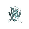
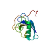
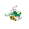
| Structure viewer | Molecule: MolmilJmol/JSmol |
|---|
- Downloads & links
-Download
| PDBx/mmCIF format | 2d5g.cif.gz | 109.4 KB | Display | PDBx/mmCIF format |
|---|---|---|---|---|
| PDB format | pdb2d5g.ent.gz | 87.4 KB | Display | PDB format |
| PDBx/mmJSON format | 2d5g.json.gz | Tree view | PDBx/mmJSON format | |
| Others |  Other downloads Other downloads |
-Validation report
| Arichive directory | https://data.pdbj.org/pub/pdb/validation_reports/d5/2d5gftp://data.pdbj.org/pub/pdb/validation_reports/d5/2d5g | HTTPS FTP |
|---|
-Related structure data
| Related structure data | |
|---|---|
| Similar structure data |


-Links
 PDBj
PDBj


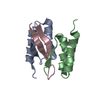
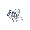
- Assembly
| Deposited unit | 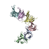
| ||||||||
|---|---|---|---|---|---|---|---|---|---|
| 1 | 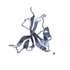
| ||||||||
| 2 | 
| ||||||||
| 3 | 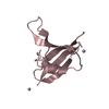
| ||||||||
| 4 | 
| ||||||||
| 5 | 
| ||||||||
| 6 | 
| ||||||||
| Unit cell |
|
-Components
| #1: Protein | Mass: 9803.242 Da / Num. of mol.: 6 / Fragment: DIX DOMAIN / Mutation: R767E Source method: isolated from a genetically manipulated source Source: (gene. exp.)  #2: Chemical | ChemComp-HG /   Mass: 200.590 Da / Num. of mol.: 10 / Source method: obtained synthetically / Formula: Hg Mass: 200.590 Da / Num. of mol.: 10 / Source method: obtained synthetically / Formula: Hg |
|---|
-Experimental details
-Experiment
| Experiment | Method: X-RAY DIFFRACTION / Number of used crystals: 1 |
|---|
- Sample preparation
| Crystal | Density Matthews: 2.98 Å3/Da / Density % sol: 58.66 % / Description: the file contains Friedel pairs. |
|---|---|
| Crystal grow | Temperature: 283 K / Method: vapor diffusion, sitting drop / pH: 7.5 Details: PEG8000, TRIS, pH 7.5, VAPOR DIFFUSION, SITTING DROP, temperature 283K |
-Data collection
| Diffraction | Mean temperature: 100 K |
|---|---|
| Diffraction source | Source: SYNCHROTRON / Site: Photon Factory  / Beamline: BL-5A / Wavelength: 1 Å / Beamline: BL-5A / Wavelength: 1 Å |
| Detector | Type: ADSC QUANTUM 4 / Detector: CCD / Date: Feb 19, 2005 |
| Radiation | Protocol: SINGLE WAVELENGTH / Monochromatic (M) / Laue (L): M / Scattering type: x-ray |
| Radiation wavelength | Wavelength: 1 Å / Relative weight: 1 |
| Reflection | Resolution: 3.2→50 Å / Num. all: 21930 / Num. obs: 21930 / % possible obs: 99.9 % / Observed criterion σ(F): 0 / Observed criterion σ(I): 0 |
| Reflection shell | Resolution: 3.2→3.31 Å / % possible all: 100 |
- Processing
| Software |
| ||||||||||||||||||||||||||||||||||||||||||||||||||||||||||||||||||||||||||||||||
|---|---|---|---|---|---|---|---|---|---|---|---|---|---|---|---|---|---|---|---|---|---|---|---|---|---|---|---|---|---|---|---|---|---|---|---|---|---|---|---|---|---|---|---|---|---|---|---|---|---|---|---|---|---|---|---|---|---|---|---|---|---|---|---|---|---|---|---|---|---|---|---|---|---|---|---|---|---|---|---|---|---|
| Refinement | Method to determine structure: MOLECULAR REPLACEMENT / Resolution: 3.2→49.18 Å / Rfactor Rfree error: 0.007 / Data cutoff high absF: 321432.46 / Data cutoff low absF: 0 / Isotropic thermal model: RESTRAINED / Cross valid method: THROUGHOUT / σ(F): 0 / Details: the file contains Friedel pairs.
| ||||||||||||||||||||||||||||||||||||||||||||||||||||||||||||||||||||||||||||||||
| Solvent computation | Solvent model: FLAT MODEL / Bsol: 66.6075 Å2 / ksol: 0.281879 e/Å3 | ||||||||||||||||||||||||||||||||||||||||||||||||||||||||||||||||||||||||||||||||
| Displacement parameters | Biso mean: 76.2 Å2
| ||||||||||||||||||||||||||||||||||||||||||||||||||||||||||||||||||||||||||||||||
| Refine analyze |
| ||||||||||||||||||||||||||||||||||||||||||||||||||||||||||||||||||||||||||||||||
| Refinement step | Cycle: LAST / Resolution: 3.2→49.18 Å
| ||||||||||||||||||||||||||||||||||||||||||||||||||||||||||||||||||||||||||||||||
| Refine LS restraints |
| ||||||||||||||||||||||||||||||||||||||||||||||||||||||||||||||||||||||||||||||||
| LS refinement shell | Resolution: 3.2→3.31 Å / Rfactor Rfree error: 0.032 / Total num. of bins used: 10
| ||||||||||||||||||||||||||||||||||||||||||||||||||||||||||||||||||||||||||||||||
| Xplor file |
|
