 Movie
Movie Controller

[English] 日本語
 Yorodumi
- PDB-2q3g: Structure of the PDZ domain of human PDLIM7 bound to a C-terminal... -
+ Open data

- Basic information
| Entry | Database: PDB / ID: 2q3g | ||||||
|---|---|---|---|---|---|---|---|
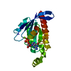
| Title | Structure of the PDZ domain of human PDLIM7 bound to a C-terminal extension from human beta-tropomyosin | ||||||
 Components Components | PDZ and LIM domain protein 7 | ||||||
 Keywords Keywords | STRUCTURAL GENOMICS / Structural Genomics Consortium / SGC | ||||||
| Function / homology |  Function and homology information Function and homology informationmuscle structure development / muscle alpha-actinin binding / filamentous actin / RET signaling / ruffle / stress fiber / receptor-mediated endocytosis / ossification / adherens junction / Z disc ...muscle structure development / muscle alpha-actinin binding / filamentous actin / RET signaling / ruffle / stress fiber / receptor-mediated endocytosis / ossification / adherens junction / Z disc / actin cytoskeleton / heart development / actin binding / actin cytoskeleton organization / cell differentiation / focal adhesion / nucleoplasm / metal ion binding / cytosol Similarity search - Function | ||||||
| Biological species |  Homo sapiens (human) Homo sapiens (human) | ||||||
| Method |  X-RAY DIFFRACTION / SYNCHROTRON / MOLECULAR REPLACEMENT / Resolution: 1.11 Å X-RAY DIFFRACTION / SYNCHROTRON / MOLECULAR REPLACEMENT / Resolution: 1.11 Å | ||||||
 Authors Authors | Gileadi, C. / Papagrigoriou, E. / Elkins, J. / Burgess-Brown, N. / Salah, E. / Gileadi, O. / Umeano, C. / Bunkoczi, G. / von Delft, F. / Uppenberg, J. ...Gileadi, C. / Papagrigoriou, E. / Elkins, J. / Burgess-Brown, N. / Salah, E. / Gileadi, O. / Umeano, C. / Bunkoczi, G. / von Delft, F. / Uppenberg, J. / Pike, A.C.W. / Arrowsmith, C.H. / Edwards, A. / Weigelt, J. / Sundstrom, M. / Doyle, D.A. / Structural Genomics Consortium (SGC) | ||||||
 Citation Citation | Journal: Protein Sci. / Year: 2010 Title: Unusual binding interactions in PDZ domain crystal structures help explain binding mechanisms Authors: Elkins, J.M. / Gileadi, C. / Shrestha, L. / Phillips, C. / Wang, J. / Muniz, J.R. / Doyle, D.A. | ||||||
| History |
|
- Structure visualization
| Structure viewer | Molecule: MolmilJmol/JSmol |
|---|
- Downloads & links
-Download
| PDBx/mmCIF format | 2q3g.cif.gz | 92.9 KB | Display | PDBx/mmCIF format |
|---|---|---|---|---|
| PDB format | pdb2q3g.ent.gz | 70.9 KB | Display | PDB format |
| PDBx/mmJSON format | 2q3g.json.gz | Tree view | PDBx/mmJSON format | |
| Others |  Other downloads Other downloads |
-Validation report
| Arichive directory | https://data.pdbj.org/pub/pdb/validation_reports/q3/2q3gftp://data.pdbj.org/pub/pdb/validation_reports/q3/2q3g | HTTPS FTP |
|---|
-Related structure data
| Related structure data | 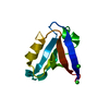 2pa1C 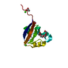 2pktC  2pntC 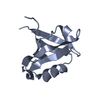 2uzcC 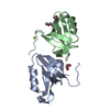 2v1wC  2w7rC C: citing same article ( |
|---|---|
| Similar structure data |


-Links
 PDBj
PDBj


- Assembly
| Deposited unit | 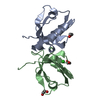
| ||||||||
|---|---|---|---|---|---|---|---|---|---|
| 1 | 
| ||||||||
| 2 | 
| ||||||||
| Unit cell |
| ||||||||
| Details | the two molecules found in the asu represent two biological units |
-Components
| #1: Protein | Mass: 9302.571 Da / Num. of mol.: 2 Source method: isolated from a genetically manipulated source Source: (gene. exp.) Homo sapiens (human) / Gene: PDLIM7 / Plasmid: pNIC28-Bsa4 / Production host:  Strain (production host): BL21(DE3) with with pRARE plasmid encoding rare codon tRNAs,(chloramphenicol-resistant) References: UniProt: Q9NR12 #2: Chemical |   Mass: 62.068 Da / Num. of mol.: 3 / Source method: obtained synthetically / Formula: C2H6O2 Mass: 62.068 Da / Num. of mol.: 3 / Source method: obtained synthetically / Formula: C2H6O2#3: Chemical | ChemComp-CL / |   Mass: 35.453 Da / Num. of mol.: 1 / Source method: obtained synthetically / Formula: Cl Mass: 35.453 Da / Num. of mol.: 1 / Source method: obtained synthetically / Formula: Cl#4: Water | ChemComp-HOH / |  Mass: 18.015 Da / Num. of mol.: 264 / Source method: isolated from a natural source / Formula: H2O Mass: 18.015 Da / Num. of mol.: 264 / Source method: isolated from a natural source / Formula: H2O |
|---|
-Experimental details
-Experiment
| Experiment | Method: X-RAY DIFFRACTION / Number of used crystals: 1 |
|---|
- Sample preparation
| Crystal | Density Matthews: 1.96 Å3/Da / Density % sol: 37.38 % |
|---|---|
| Crystal grow | Temperature: 298 K / Method: vapor diffusion, sitting drop / pH: 7.5 Details: 0.1M HEPES; 10% isopropanol; 20% PEG 4K, pH 7.5, VAPOR DIFFUSION, SITTING DROP, temperature 298K |
-Data collection
| Diffraction | Mean temperature: 100 K |
|---|---|
| Diffraction source | Source: SYNCHROTRON / Site: SLS  / Beamline: X10SA / Wavelength: 0.9182 Å / Beamline: X10SA / Wavelength: 0.9182 Å |
| Detector | Type: MARMOSAIC 225 mm CCD / Detector: CCD / Date: Feb 11, 2007 |
| Radiation | Monochromator: Si(111) / Protocol: SINGLE WAVELENGTH / Monochromatic (M) / Laue (L): M / Scattering type: x-ray |
| Radiation wavelength | Wavelength: 0.9182 Å / Relative weight: 1 |
| Reflection | Resolution: 1.11→39.84 Å / Num. all: 58681 / Num. obs: 58329 / % possible obs: 99.4 % / Observed criterion σ(F): 0 / Observed criterion σ(I): 0 / Redundancy: 5.1 % / Rmerge(I) obs: 0.042 / Rsym value: 0.038 |
| Reflection shell | Resolution: 1.11→1.15 Å / Redundancy: 4.6 % / Rmerge(I) obs: 0.144 / Mean I/σ(I) obs: 11 / Rsym value: 0.121 / % possible all: 100 |
- Processing
| Software |
| ||||||||||||||||||||||||||||||||||||||||||||||||||||||||||||||||||||||||||||||||||||||||||||||||||||||||||||||||||||||||||||||||||||||||||||||||||||||||||||||||||||||||||
|---|---|---|---|---|---|---|---|---|---|---|---|---|---|---|---|---|---|---|---|---|---|---|---|---|---|---|---|---|---|---|---|---|---|---|---|---|---|---|---|---|---|---|---|---|---|---|---|---|---|---|---|---|---|---|---|---|---|---|---|---|---|---|---|---|---|---|---|---|---|---|---|---|---|---|---|---|---|---|---|---|---|---|---|---|---|---|---|---|---|---|---|---|---|---|---|---|---|---|---|---|---|---|---|---|---|---|---|---|---|---|---|---|---|---|---|---|---|---|---|---|---|---|---|---|---|---|---|---|---|---|---|---|---|---|---|---|---|---|---|---|---|---|---|---|---|---|---|---|---|---|---|---|---|---|---|---|---|---|---|---|---|---|---|---|---|---|---|---|---|---|---|
| Refinement | Method to determine structure: MOLECULAR REPLACEMENT Starting model: homoly based model Resolution: 1.11→39.84 Å / Cor.coef. Fo:Fc: 0.977 / Cor.coef. Fo:Fc free: 0.97 / SU B: 0.657 / SU ML: 0.015 / Cross valid method: THROUGHOUT / σ(F): 0 / σ(I): 0 / ESU R: 0.031 / ESU R Free: 0.031 / Stereochemistry target values: MAXIMUM LIKELIHOOD / Details: HYDROGENS HAVE BEEN ADDED IN THE RIDING POSITIONS
| ||||||||||||||||||||||||||||||||||||||||||||||||||||||||||||||||||||||||||||||||||||||||||||||||||||||||||||||||||||||||||||||||||||||||||||||||||||||||||||||||||||||||||
| Solvent computation | Ion probe radii: 0.8 Å / Shrinkage radii: 0.8 Å / VDW probe radii: 1.4 Å / Solvent model: BABINET MODEL WITH MASK | ||||||||||||||||||||||||||||||||||||||||||||||||||||||||||||||||||||||||||||||||||||||||||||||||||||||||||||||||||||||||||||||||||||||||||||||||||||||||||||||||||||||||||
| Displacement parameters | Biso mean: 7.247 Å2
| ||||||||||||||||||||||||||||||||||||||||||||||||||||||||||||||||||||||||||||||||||||||||||||||||||||||||||||||||||||||||||||||||||||||||||||||||||||||||||||||||||||||||||
| Refinement step | Cycle: LAST / Resolution: 1.11→39.84 Å
| ||||||||||||||||||||||||||||||||||||||||||||||||||||||||||||||||||||||||||||||||||||||||||||||||||||||||||||||||||||||||||||||||||||||||||||||||||||||||||||||||||||||||||
| Refine LS restraints |
| ||||||||||||||||||||||||||||||||||||||||||||||||||||||||||||||||||||||||||||||||||||||||||||||||||||||||||||||||||||||||||||||||||||||||||||||||||||||||||||||||||||||||||
| LS refinement shell | Resolution: 1.11→1.138 Å / Total num. of bins used: 20
|
