 Movie
Movie Controller

[English] 日本語
 Yorodumi
- PDB-6rrk: Crystal structure of the central region of human cohesin subunit ... -
+ Open data

- Basic information
| Entry | Database: PDB / ID: 6rrk | ||||||
|---|---|---|---|---|---|---|---|
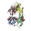
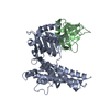
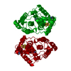
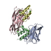
| Title | Crystal structure of the central region of human cohesin subunit STAG1 in complex with RAD21 peptide | ||||||
 Components Components |
| ||||||
 Keywords Keywords | GENE REGULATION / cohesin / Stromal antigen / chromatid | ||||||
| Function / homology |  Function and homology information Function and homology informationnegative regulation of mitotic metaphase/anaphase transition / Cohesin Loading onto Chromatin / meiotic cohesin complex / Establishment of Sister Chromatid Cohesion / establishment of meiotic sister chromatid cohesion / cohesin complex / mitotic cohesin complex / positive regulation of sister chromatid cohesion / negative regulation of glial cell apoptotic process / negative regulation of G2/M transition of mitotic cell cycle ...negative regulation of mitotic metaphase/anaphase transition / Cohesin Loading onto Chromatin / meiotic cohesin complex / Establishment of Sister Chromatid Cohesion / establishment of meiotic sister chromatid cohesion / cohesin complex / mitotic cohesin complex / positive regulation of sister chromatid cohesion / negative regulation of glial cell apoptotic process / negative regulation of G2/M transition of mitotic cell cycle / establishment of mitotic sister chromatid cohesion / replication-born double-strand break repair via sister chromatid exchange / chromatin looping / reciprocal meiotic recombination / negative regulation of interleukin-1 beta production / sister chromatid cohesion / mitotic spindle pole / lncRNA binding / positive regulation of interleukin-10 production / negative regulation of tumor necrosis factor production / mitotic spindle assembly / chromosome, centromeric region / SUMOylation of DNA damage response and repair proteins / cis-regulatory region sequence-specific DNA binding / protein localization to chromatin / Meiotic synapsis / Resolution of Sister Chromatid Cohesion / condensed nuclear chromosome / chromosome segregation / nuclear matrix / spindle pole / Separation of Sister Chromatids / double-strand break repair / chromosome / DNA recombination / midbody / Estrogen-dependent gene expression / DNA-binding transcription factor binding / negative regulation of neuron apoptotic process / response to hypoxia / cilium / nuclear body / cell division / apoptotic process / chromatin binding / regulation of transcription by RNA polymerase II / chromatin / nucleoplasm / membrane / nucleus / cytosol Similarity search - Function | ||||||
| Biological species |  Homo sapiens (human) Homo sapiens (human) | ||||||
| Method |  X-RAY DIFFRACTION / SYNCHROTRON / MOLECULAR REPLACEMENT / Resolution: 3.17 Å X-RAY DIFFRACTION / SYNCHROTRON / MOLECULAR REPLACEMENT / Resolution: 3.17 Å | ||||||
 Authors Authors | Newman, J.A. / katis, V.L. / von Delft, F. / Arrowsmith, C.H. / Edwards, A. / Bountra, C. / Gileadi, O. | ||||||
| Funding support |  United Kingdom, 1items United Kingdom, 1items
| ||||||
 Citation Citation | Journal: Life Sci Alliance / Year: 2020 Title: STAG1 vulnerabilities for exploiting cohesin synthetic lethality in STAG2-deficient cancers. Authors: van der Lelij, P. / Newman, J.A. / Lieb, S. / Jude, J. / Katis, V. / Hoffmann, T. / Hinterndorfer, M. / Bader, G. / Kraut, N. / Pearson, M.A. / Peters, J.M. / Zuber, J. / Gileadi, O. / Petronczki, M. | ||||||
| History |
|
- Structure visualization
| Structure viewer | Molecule: MolmilJmol/JSmol |
|---|
- Downloads & links
-Download
| PDBx/mmCIF format | 6rrk.cif.gz | 191.5 KB | Display | PDBx/mmCIF format |
|---|---|---|---|---|
| PDB format | pdb6rrk.ent.gz | 151.9 KB | Display | PDB format |
| PDBx/mmJSON format | 6rrk.json.gz | Tree view | PDBx/mmJSON format | |
| Others |  Other downloads Other downloads |
-Validation report
| Arichive directory | https://data.pdbj.org/pub/pdb/validation_reports/rr/6rrkftp://data.pdbj.org/pub/pdb/validation_reports/rr/6rrk | HTTPS FTP |
|---|
-Related structure data
| Related structure data |  6qb5C  6r7oC  6rrcC  4pk7S S: Starting model for refinement C: citing same article ( |
|---|---|
| Similar structure data |


-Links
 PDBj
PDBj


- Assembly
| Deposited unit | 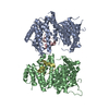
| ||||||||
|---|---|---|---|---|---|---|---|---|---|
| 1 | 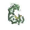
| ||||||||
| 2 | 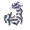
| ||||||||
| Unit cell |
|
-Components
| #1: Protein | Mass: 52752.941 Da / Num. of mol.: 2 Source method: isolated from a genetically manipulated source Source: (gene. exp.) Homo sapiens (human) / Gene: STAG1, SA1, SCC3 / Production host:  #2: Protein/peptide | Mass: 4694.692 Da / Num. of mol.: 2 / Source method: obtained synthetically / Source: (synth.) Homo sapiens (human) / References: UniProt: O60216 |
|---|
-Experimental details
-Experiment
| Experiment | Method: X-RAY DIFFRACTION / Number of used crystals: 1 |
|---|
- Sample preparation
| Crystal | Density Matthews: 3.6 Å3/Da / Density % sol: 65.82 % |
|---|---|
| Crystal grow | Temperature: 277 K / Method: vapor diffusion, sitting drop / Details: 16 % PEG 3350, 0.2 M DL-Mallic acid |
-Data collection
| Diffraction | Mean temperature: 100 K / Serial crystal experiment: N |
|---|---|
| Diffraction source | Source: SYNCHROTRON / Site: Diamond / Beamline: I24 / Wavelength: 0.968 Å |
| Detector | Type: DECTRIS PILATUS3 6M / Detector: PIXEL / Date: Oct 22, 2017 |
| Radiation | Protocol: SINGLE WAVELENGTH / Monochromatic (M) / Laue (L): M / Scattering type: x-ray |
| Radiation wavelength | Wavelength: 0.968 Å / Relative weight: 1 |
| Reflection | Resolution: 3.17→69.67 Å / Num. obs: 24764 / % possible obs: 97 % / Redundancy: 2.8 % / CC1/2: 0.99 / Rmerge(I) obs: 0.066 / Rpim(I) all: 0.046 / Net I/σ(I): 7.3 |
| Reflection shell | Resolution: 3.17→3.39 Å / Redundancy: 2.8 % / Rmerge(I) obs: 0.391 / Mean I/σ(I) obs: 1.9 / Num. unique obs: 4500 / CC1/2: 0.914 / Rpim(I) all: 0.265 / % possible all: 97.8 |
- Processing
| Software |
| |||||||||||||||||||||||||||||||||||||||||||||||||||||||||||||||
|---|---|---|---|---|---|---|---|---|---|---|---|---|---|---|---|---|---|---|---|---|---|---|---|---|---|---|---|---|---|---|---|---|---|---|---|---|---|---|---|---|---|---|---|---|---|---|---|---|---|---|---|---|---|---|---|---|---|---|---|---|---|---|---|---|
| Refinement | Method to determine structure: MOLECULAR REPLACEMENT Starting model: 4pk7 Resolution: 3.17→60.229 Å / SU ML: 0.53 / Cross valid method: FREE R-VALUE / σ(F): 1.38 / Phase error: 36.93 / Stereochemistry target values: ML
| |||||||||||||||||||||||||||||||||||||||||||||||||||||||||||||||
| Solvent computation | Shrinkage radii: 0.9 Å / VDW probe radii: 1.11 Å / Solvent model: FLAT BULK SOLVENT MODEL | |||||||||||||||||||||||||||||||||||||||||||||||||||||||||||||||
| Refinement step | Cycle: LAST / Resolution: 3.17→60.229 Å
| |||||||||||||||||||||||||||||||||||||||||||||||||||||||||||||||
| Refine LS restraints |
| |||||||||||||||||||||||||||||||||||||||||||||||||||||||||||||||
| LS refinement shell |
|
