- PDB-4rg8: Structural and biochemical studies of a moderately thermophilic E... -
+
Open data
ID or keywords:
Loading...
-
Basic information
Entry
Database: PDB / ID: 4rg8
Title
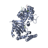
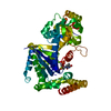
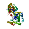
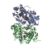
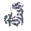
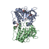
Structural and biochemical studies of a moderately thermophilic Exonuclease I from Methylocaldum szegediense
Components
Exonuclease I
Keywords
HYDROLASE / Methylocaldum szegediense / Exonuclease / DNA binding protein
Function / homology
Function and homology information
exodeoxyribonuclease I / single-stranded DNA 3'-5' DNA exonuclease activity / 3'-5'-RNA exonuclease activity / DNA repair / DNA binding / metal ion binding Similarity search - Function
Exonuclease I, C-terminal alpha-helical domain / Helix Hairpins - #1240 / Exonuclease ExoI, domain 3 / Exonuclease ExoI, domain 2 / Exodeoxyribonuclease I, C-terminal / Exodeoxyribonuclease I / Exonuclease I, SH3-like domain / Exonuclease I, SH3-like domain superfamily / Exonuclease I SH3-like domain / Exonuclease I (ExoI) SH3-like domain profile. ...Exonuclease I, C-terminal alpha-helical domain / Helix Hairpins - #1240 / Exonuclease ExoI, domain 3 / Exonuclease ExoI, domain 2 / Exodeoxyribonuclease I, C-terminal / Exodeoxyribonuclease I / Exonuclease I, SH3-like domain / Exonuclease I, SH3-like domain superfamily / Exonuclease I SH3-like domain / Exonuclease I (ExoI) SH3-like domain profile. / Exonuclease I (ExoI) C-terminal domain profile. / Oligoribonuclease / PX Domain / Exonuclease / Exonuclease, RNase T/DNA polymerase III / EXOIII / Monooxygenase / Ribonuclease H-like superfamily/Ribonuclease H / Nucleotidyltransferase; domain 5 / Helix Hairpins / Ribonuclease H superfamily / Ribonuclease H-like superfamily / Up-down Bundle / 2-Layer Sandwich / Orthogonal Bundle / Mainly Alpha / Alpha Beta Similarity search - Domain/homology
Mass: 18.015 Da / Num. of mol.: 189 / Source method: isolated from a natural source / Formula: H2O
-
Experimental details
-
Experiment
Experiment
Method: X-RAY DIFFRACTION / Number of used crystals: 1
-
Sample preparation
Crystal
Density Matthews: 2.78 Å3/Da / Density % sol: 55.75 %
Crystal grow
Temperature: 298 K / Method: vapor diffusion, sitting drop / pH: 8 Details: 20% PEG6K, 0.1 M Tris pH8, 0.2 M calcium chloride , VAPOR DIFFUSION, SITTING DROP, temperature 298K
Resolution: 2.12→29.08 Å / Cor.coef. Fo:Fc: 0.949 / Cor.coef. Fo:Fc free: 0.93 / SU B: 6.618 / SU ML: 0.163 / Cross valid method: THROUGHOUT / ESU R: 0.214 / ESU R Free: 0.181 / Stereochemistry target values: MAXIMUM LIKELIHOOD / Details: HYDROGENS HAVE BEEN ADDED IN THE RIDING POSITIONS
Rfactor
Num. reflection
% reflection
Selection details
Rfree
0.25292
1912
5 %
RANDOM
Rwork
0.22278
-
-
-
all
0.22278
-
-
-
obs
0.22433
36277
99.22 %
-
Solvent computation
Ion probe radii: 0.8 Å / Shrinkage radii: 0.8 Å / VDW probe radii: 1.2 Å / Solvent model: MASK
Displacement parameters
Biso mean: 49.365 Å2
Baniso -1
Baniso -2
Baniso -3
1-
-0.34 Å2
-0 Å2
-0 Å2
2-
-
0.44 Å2
-0 Å2
3-
-
-
-0.1 Å2
Refinement step
Cycle: LAST / Resolution: 2.12→29.08 Å
Protein
Nucleic acid
Ligand
Solvent
Total
Num. atoms
3768
0
1
189
3958
Refine LS restraints
Refine-ID
Type
Dev ideal
Dev ideal target
Number
X-RAY DIFFRACTION
r_bond_refined_d
0.005
0.019
3910
X-RAY DIFFRACTION
r_bond_other_d
0.001
0.02
3772
X-RAY DIFFRACTION
r_angle_refined_deg
1.029
1.976
5318
X-RAY DIFFRACTION
r_angle_other_deg
0.741
3
8653
X-RAY DIFFRACTION
r_dihedral_angle_1_deg
5.324
5
480
X-RAY DIFFRACTION
r_dihedral_angle_2_deg
33.702
22.338
201
X-RAY DIFFRACTION
r_dihedral_angle_3_deg
14.626
15
672
X-RAY DIFFRACTION
r_dihedral_angle_4_deg
14.239
15
54
X-RAY DIFFRACTION
r_chiral_restr
0.059
0.2
584
X-RAY DIFFRACTION
r_gen_planes_refined
0.004
0.021
4420
X-RAY DIFFRACTION
r_gen_planes_other
0.001
0.02
926
X-RAY DIFFRACTION
r_mcbond_it
1.555
4.861
1884
X-RAY DIFFRACTION
r_mcbond_other
1.555
4.86
1883
X-RAY DIFFRACTION
r_mcangle_it
2.624
7.279
2355
X-RAY DIFFRACTION
r_mcangle_other
2.623
7.281
2356
X-RAY DIFFRACTION
r_scbond_it
1.596
5.049
2026
X-RAY DIFFRACTION
r_scbond_other
1.596
5.049
2026
X-RAY DIFFRACTION
r_scangle_other
2.755
7.498
2957
X-RAY DIFFRACTION
r_long_range_B_refined
4.959
38.154
4530
X-RAY DIFFRACTION
r_long_range_B_other
4.804
38.115
4479
LS refinement shell
Resolution: 2.115→2.17 Å / Total num. of bins used: 20
Rfactor
Num. reflection
% reflection
Rfree
0.353
128
-
Rwork
0.336
2523
-
obs
-
-
94.92 %
+
About Yorodumi
-
News
-
Feb 9, 2022. New format data for meta-information of EMDB entries
New format data for meta-information of EMDB entries
Version 3 of the EMDB header file is now the official format.
The previous official version 1.9 will be removed from the archive.
In the structure databanks used in Yorodumi, some data are registered as the other names, "COVID-19 virus" and "2019-nCoV". Here are the details of the virus and the list of structure data.
Jan 31, 2019. EMDB accession codes are about to change! (news from PDBe EMDB page)
EMDB accession codes are about to change! (news from PDBe EMDB page)
The allocation of 4 digits for EMDB accession codes will soon come to an end. Whilst these codes will remain in use, new EMDB accession codes will include an additional digit and will expand incrementally as the available range of codes is exhausted. The current 4-digit format prefixed with “EMD-” (i.e. EMD-XXXX) will advance to a 5-digit format (i.e. EMD-XXXXX), and so on. It is currently estimated that the 4-digit codes will be depleted around Spring 2019, at which point the 5-digit format will come into force.
The EM Navigator/Yorodumi systems omit the EMD- prefix.
Related info.:Q: What is EMD? / ID/Accession-code notation in Yorodumi/EM Navigator
Yorodumi is a browser for structure data from EMDB, PDB, SASBDB, etc.
This page is also the successor to EM Navigator detail page, and also detail information page/front-end page for Omokage search.
The word "yorodu" (or yorozu) is an old Japanese word meaning "ten thousand". "mi" (miru) is to see.
Related info.:EMDB / PDB / SASBDB / Comparison of 3 databanks / Yorodumi Search / Aug 31, 2016. New EM Navigator & Yorodumi / Yorodumi Papers / Jmol/JSmol / Function and homology information / Changes in new EM Navigator and Yorodumi
 Movie
Movie Controller

 Yorodumi
 Open data
 Basic information
 Components
 Keywords
 Function and homology information
 Methylocaldum szegediense (bacteria)
 X-RAY DIFFRACTION /
  Authors
 Citation
 Structure visualization
 Downloads & links
 Other downloads
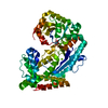


 PDBj
PDBj Assembly

 Mass: 24.305 Da / Num. of mol.: 1 / Source method: obtained synthetically / Formula: Mg
 Mass: 18.015 Da / Num. of mol.: 189 / Source method: isolated from a natural source / Formula: H2O
 Sample preparation
 / Beamline: I02 / Wavelength: 0.9795 Å
 Processing