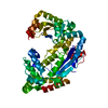
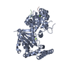
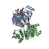
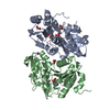
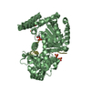
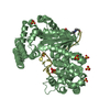
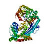
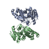
- PDB-3hl8: Crystal structure of exonuclease I in complex with inhibitor BCBP -
+
Open data
ID or keywords:
Loading...
-
Basic information
Entry
Database: PDB / ID: 3hl8
Title
Crystal structure of exonuclease I in complex with inhibitor BCBP
Components
Exodeoxyribonuclease I
Keywords
HYDROLASE / EXONUCLEASE / SSB / GENOME MAINTENANCE / DNA DAMAGE / DNA REPAIR / Nuclease
Function / homology
Function and homology information
exodeoxyribonuclease I / DNA replication termination / single-stranded DNA 3'-5' DNA exonuclease activity / DNA catabolic process / 5'-deoxyribose-5-phosphate lyase activity / single-stranded DNA binding / 3'-5'-RNA exonuclease activity / DNA repair / magnesium ion binding Similarity search - Function
: / Exonuclease I C-terminal domain / Helix Hairpins - #1240 / Exonuclease ExoI, domain 3 / Exonuclease ExoI, domain 2 / Exodeoxyribonuclease I, C-terminal / Exodeoxyribonuclease I / Exonuclease I, SH3-like domain / Exonuclease I, SH3-like domain superfamily / Exonuclease I SH3-like domain ...: / Exonuclease I C-terminal domain / Helix Hairpins - #1240 / Exonuclease ExoI, domain 3 / Exonuclease ExoI, domain 2 / Exodeoxyribonuclease I, C-terminal / Exodeoxyribonuclease I / Exonuclease I, SH3-like domain / Exonuclease I, SH3-like domain superfamily / Exonuclease I SH3-like domain / Exonuclease I (ExoI) SH3-like domain profile. / Exonuclease I (ExoI) C-terminal domain profile. / Oligoribonuclease / PX Domain / Exonuclease / Exonuclease, RNase T/DNA polymerase III / EXOIII / Monooxygenase / Ribonuclease H-like superfamily/Ribonuclease H / Nucleotidyltransferase; domain 5 / Helix Hairpins / Ribonuclease H superfamily / Ribonuclease H-like superfamily / Up-down Bundle / 2-Layer Sandwich / Orthogonal Bundle / Mainly Alpha / Alpha Beta Similarity search - Domain/homology
In the structure databanks used in Yorodumi, some data are registered as the other names, "COVID-19 virus" and "2019-nCoV". Here are the details of the virus and the list of structure data.
Jan 31, 2019. EMDB accession codes are about to change! (news from PDBe EMDB page)
EMDB accession codes are about to change! (news from PDBe EMDB page)
The allocation of 4 digits for EMDB accession codes will soon come to an end. Whilst these codes will remain in use, new EMDB accession codes will include an additional digit and will expand incrementally as the available range of codes is exhausted. The current 4-digit format prefixed with “EMD-” (i.e. EMD-XXXX) will advance to a 5-digit format (i.e. EMD-XXXXX), and so on. It is currently estimated that the 4-digit codes will be depleted around Spring 2019, at which point the 5-digit format will come into force.
The EM Navigator/Yorodumi systems omit the EMD- prefix.
Related info.:Q: What is EMD? / ID/Accession-code notation in Yorodumi/EM Navigator
Yorodumi is a browser for structure data from EMDB, PDB, SASBDB, etc.
This page is also the successor to EM Navigator detail page, and also detail information page/front-end page for Omokage search.
The word "yorodu" (or yorozu) is an old Japanese word meaning "ten thousand". "mi" (miru) is to see.
Related info.:EMDB / PDB / SASBDB / Comparison of 3 databanks / Yorodumi Search / Aug 31, 2016. New EM Navigator & Yorodumi / Yorodumi Papers / Jmol/JSmol / Function and homology information / Changes in new EM Navigator and Yorodumi
 Movie
Movie Controller

 Yorodumi
 Open data
 Basic information
 Components
 Keywords
 Function and homology information
 X-RAY DIFFRACTION /
  Authors
 Citation
 Structure visualization
 Downloads & links
 Other downloads
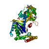


 PDBj
PDBj Assembly
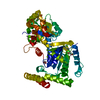


 Mass: 24.305 Da / Num. of mol.: 1 / Source method: obtained synthetically / Formula: Mg
 Mass: 307.798 Da / Num. of mol.: 1 / Source method: obtained synthetically / Formula: C14H14ClN3OS
 Mass: 78.133 Da / Num. of mol.: 2 / Source method: obtained synthetically / Formula: C2H6OS / Comment: DMSO, precipitant*YM
 Mass: 62.068 Da / Num. of mol.: 3 / Source method: obtained synthetically / Formula: C2H6O2
 Sample preparation
 / Beamline: 21-ID-D / Wavelength: 0.97869 Å
 Processing