 Movie
Movie Controller

[English] 日本語
 Yorodumi
- PDB-6qb5: Crystal structure of the N-terminal region of human cohesin subun... -
+ Open data

- Basic information
| Entry | Database: PDB / ID: 6qb5 | ||||||
|---|---|---|---|---|---|---|---|
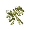
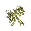
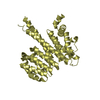
| Title | Crystal structure of the N-terminal region of human cohesin subunit STAG1 | ||||||
 Components Components | Cohesin subunit SA-1 | ||||||
 Keywords Keywords | GENE REGULATION / Cohesin / SA-1 / chromosome segregation | ||||||
| Function / homology |  Function and homology information Function and homology informationCohesin Loading onto Chromatin / Establishment of Sister Chromatid Cohesion / cohesin complex / mitotic cohesin complex / establishment of mitotic sister chromatid cohesion / sister chromatid cohesion / mitotic spindle pole / mitotic spindle assembly / chromosome, centromeric region / SUMOylation of DNA damage response and repair proteins ...Cohesin Loading onto Chromatin / Establishment of Sister Chromatid Cohesion / cohesin complex / mitotic cohesin complex / establishment of mitotic sister chromatid cohesion / sister chromatid cohesion / mitotic spindle pole / mitotic spindle assembly / chromosome, centromeric region / SUMOylation of DNA damage response and repair proteins / Meiotic synapsis / Resolution of Sister Chromatid Cohesion / nuclear matrix / Separation of Sister Chromatids / chromosome / Estrogen-dependent gene expression / cilium / nuclear body / cell division / chromatin binding / chromatin / nucleoplasm / nucleus / cytosol Similarity search - Function | ||||||
| Biological species |  Homo sapiens (human) Homo sapiens (human) | ||||||
| Method |  X-RAY DIFFRACTION / SYNCHROTRON / MOLECULAR REPLACEMENT / Resolution: 2.02 Å X-RAY DIFFRACTION / SYNCHROTRON / MOLECULAR REPLACEMENT / Resolution: 2.02 Å | ||||||
 Authors Authors | Newman, J.A. / Katis, V.L. / von Delft, F. / Arrowsmith, C.H. / Edwards, A. / Bountra, C. / Gileadi, O. | ||||||
| Funding support | 1items
| ||||||
 Citation Citation | Journal: Life Sci Alliance / Year: 2020 Title: STAG1 vulnerabilities for exploiting cohesin synthetic lethality in STAG2-deficient cancers. Authors: van der Lelij, P. / Newman, J.A. / Lieb, S. / Jude, J. / Katis, V. / Hoffmann, T. / Hinterndorfer, M. / Bader, G. / Kraut, N. / Pearson, M.A. / Peters, J.M. / Zuber, J. / Gileadi, O. / Petronczki, M. | ||||||
| History |
|
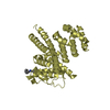
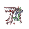
- Structure visualization
| Structure viewer | Molecule: MolmilJmol/JSmol |
|---|
- Downloads & links
-Download
| PDBx/mmCIF format | 6qb5.cif.gz | 270.4 KB | Display | PDBx/mmCIF format |
|---|---|---|---|---|
| PDB format | pdb6qb5.ent.gz | 215.8 KB | Display | PDB format |
| PDBx/mmJSON format | 6qb5.json.gz | Tree view | PDBx/mmJSON format | |
| Others |  Other downloads Other downloads |
-Validation report
| Arichive directory | https://data.pdbj.org/pub/pdb/validation_reports/qb/6qb5ftp://data.pdbj.org/pub/pdb/validation_reports/qb/6qb5 | HTTPS FTP |
|---|
-Related structure data
| Related structure data |  6r7oC  6rrcC  6rrkC  4pk7S S: Starting model for refinement C: citing same article ( |
|---|---|
| Similar structure data |

-Links
 PDBj
PDBj


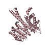
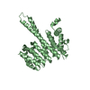
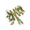
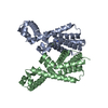
- Assembly
| Deposited unit | 
| ||||||||
|---|---|---|---|---|---|---|---|---|---|
| 1 | 
| ||||||||
| 2 | 
| ||||||||
| 3 | 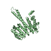
| ||||||||
| 4 | 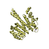
| ||||||||
| Unit cell |
|
-Components
| #1: Protein | Mass: 39629.531 Da / Num. of mol.: 4 Source method: isolated from a genetically manipulated source Source: (gene. exp.) Homo sapiens (human) / Gene: STAG1, SA1, SCC3 / Production host:  #2: Chemical | ChemComp-NA / |   Mass: 22.990 Da / Num. of mol.: 1 / Source method: obtained synthetically / Formula: Na Mass: 22.990 Da / Num. of mol.: 1 / Source method: obtained synthetically / Formula: Na#3: Water | ChemComp-HOH / |  Mass: 18.015 Da / Num. of mol.: 627 / Source method: isolated from a natural source / Formula: H2O Mass: 18.015 Da / Num. of mol.: 627 / Source method: isolated from a natural source / Formula: H2O |
|---|
-Experimental details
-Experiment
| Experiment | Method: X-RAY DIFFRACTION / Number of used crystals: 1 |
|---|
- Sample preparation
| Crystal | Density Matthews: 3 Å3/Da / Density % sol: 58.99 % |
|---|---|
| Crystal grow | Temperature: 277 K / Method: vapor diffusion, sitting drop Details: 0.1 M Na/K Phosphate pH 6.0, 0.2 M NaCl, 34% PEG200 |
-Data collection
| Diffraction | Mean temperature: 100 K / Serial crystal experiment: N |
|---|---|
| Diffraction source | Source: SYNCHROTRON / Site: Diamond  / Beamline: I04-1 / Wavelength: 0.9159 Å / Beamline: I04-1 / Wavelength: 0.9159 Å |
| Detector | Type: DECTRIS PILATUS3 6M / Detector: PIXEL / Date: May 27, 2018 |
| Radiation | Protocol: SINGLE WAVELENGTH / Monochromatic (M) / Laue (L): M / Scattering type: x-ray |
| Radiation wavelength | Wavelength: 0.9159 Å / Relative weight: 1 |
| Reflection | Resolution: 2.02→110 Å / Num. obs: 125560 / % possible obs: 99.8 % / Redundancy: 6 % / CC1/2: 0.998 / Rmerge(I) obs: 0.081 / Rpim(I) all: 0.052 / Net I/σ(I): 9.8 |
| Reflection shell | Resolution: 2.02→2.07 Å / Redundancy: 4.4 % / Rmerge(I) obs: 0.969 / Mean I/σ(I) obs: 1.1 / Num. unique obs: 9012 / CC1/2: 0.664 / Rpim(I) all: 0.583 / % possible all: 98 |
- Processing
| Software |
| |||||||||||||||||||||||||||||||||||||||||||||||||||||||||||||||||||||||||||||||||||||||||||||||||||||||||||||||||||||||||||||||||||||||||||||||||||||||||||||||||||||||||||||||||||||||||||||||||||||||||||||||||||||||||
|---|---|---|---|---|---|---|---|---|---|---|---|---|---|---|---|---|---|---|---|---|---|---|---|---|---|---|---|---|---|---|---|---|---|---|---|---|---|---|---|---|---|---|---|---|---|---|---|---|---|---|---|---|---|---|---|---|---|---|---|---|---|---|---|---|---|---|---|---|---|---|---|---|---|---|---|---|---|---|---|---|---|---|---|---|---|---|---|---|---|---|---|---|---|---|---|---|---|---|---|---|---|---|---|---|---|---|---|---|---|---|---|---|---|---|---|---|---|---|---|---|---|---|---|---|---|---|---|---|---|---|---|---|---|---|---|---|---|---|---|---|---|---|---|---|---|---|---|---|---|---|---|---|---|---|---|---|---|---|---|---|---|---|---|---|---|---|---|---|---|---|---|---|---|---|---|---|---|---|---|---|---|---|---|---|---|---|---|---|---|---|---|---|---|---|---|---|---|---|---|---|---|---|---|---|---|---|---|---|---|---|---|---|---|---|---|---|---|---|
| Refinement | Method to determine structure: MOLECULAR REPLACEMENT Starting model: 4pk7 Resolution: 2.02→85.002 Å / SU ML: 0.26 / Cross valid method: FREE R-VALUE / σ(F): 1.34 / Phase error: 28.54
| |||||||||||||||||||||||||||||||||||||||||||||||||||||||||||||||||||||||||||||||||||||||||||||||||||||||||||||||||||||||||||||||||||||||||||||||||||||||||||||||||||||||||||||||||||||||||||||||||||||||||||||||||||||||||
| Solvent computation | Shrinkage radii: 0.9 Å / VDW probe radii: 1.11 Å | |||||||||||||||||||||||||||||||||||||||||||||||||||||||||||||||||||||||||||||||||||||||||||||||||||||||||||||||||||||||||||||||||||||||||||||||||||||||||||||||||||||||||||||||||||||||||||||||||||||||||||||||||||||||||
| Refinement step | Cycle: LAST / Resolution: 2.02→85.002 Å
| |||||||||||||||||||||||||||||||||||||||||||||||||||||||||||||||||||||||||||||||||||||||||||||||||||||||||||||||||||||||||||||||||||||||||||||||||||||||||||||||||||||||||||||||||||||||||||||||||||||||||||||||||||||||||
| Refine LS restraints |
| |||||||||||||||||||||||||||||||||||||||||||||||||||||||||||||||||||||||||||||||||||||||||||||||||||||||||||||||||||||||||||||||||||||||||||||||||||||||||||||||||||||||||||||||||||||||||||||||||||||||||||||||||||||||||
| LS refinement shell |
|
