 Movie
Movie Controller

[English] 日本語
 Yorodumi
- PDB-6rpr: LEM domain of Emerin mutant T43I in complex with BAF dimer and th... -
+ Open data

- Basic information
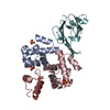
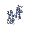
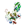
| Entry | Database: PDB / ID: 6rpr | |||||||||
|---|---|---|---|---|---|---|---|---|---|---|
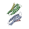
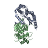
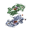
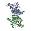
| Title | LEM domain of Emerin mutant T43I in complex with BAF dimer and the Igfold of the lamin A/C | |||||||||
 Components Components |
| |||||||||
 Keywords Keywords | DNA BINDING PROTEIN / Nuclear membrane protein | |||||||||
| Function / homology |  Function and homology information Function and homology informationTMEM240-body / nuclear membrane organization / structural constituent of nuclear lamina / negative regulation of protein ADP-ribosylation / protein localization to nuclear envelope / establishment or maintenance of microtubule cytoskeleton polarity / Breakdown of the nuclear lamina / Depolymerization of the Nuclear Lamina / nuclear envelope organization / Nuclear Envelope Breakdown ...TMEM240-body / nuclear membrane organization / structural constituent of nuclear lamina / negative regulation of protein ADP-ribosylation / protein localization to nuclear envelope / establishment or maintenance of microtubule cytoskeleton polarity / Breakdown of the nuclear lamina / Depolymerization of the Nuclear Lamina / nuclear envelope organization / Nuclear Envelope Breakdown / nuclear pore localization / DNA double-strand break attachment to nuclear envelope / lamin filament / mitotic nuclear membrane reassembly / XBP1(S) activates chaperone genes / nuclear lamina / Initiation of Nuclear Envelope (NE) Reformation / RHOD GTPase cycle / Integration of viral DNA into host genomic DNA / Autointegration results in viral DNA circles / regulation of canonical Wnt signaling pathway / nuclear inner membrane / regulation of telomere maintenance / intermediate filament / nuclear migration / negative regulation of cardiac muscle hypertrophy in response to stress / muscle organ development / Insertion of tail-anchored proteins into the endoplasmic reticulum membrane / negative regulation of viral genome replication / 2-LTR circle formation / negative regulation of cGAS/STING signaling pathway / Vpr-mediated nuclear import of PICs / Deregulated CDK5 triggers multiple neurodegenerative pathways in Alzheimer's disease models / negative regulation of type I interferon production / Integration of provirus / APOBEC3G mediated resistance to HIV-1 infection / skeletal muscle cell differentiation / nuclear outer membrane / RHOG GTPase cycle / RAC3 GTPase cycle / RAC2 GTPase cycle / chromosome organization / negative regulation of fibroblast proliferation / protein localization to nucleus / beta-tubulin binding / condensed chromosome / RAC1 GTPase cycle / muscle contraction / regulation of cell migration / negative regulation of innate immune response / positive regulation of protein export from nucleus / Meiotic synapsis / negative regulation of canonical Wnt signaling pathway / cellular response to growth factor stimulus / response to virus / double-strand break repair via nonhomologous end joining / cellular senescence / spindle / structural constituent of cytoskeleton / nuclear matrix / intracellular protein localization / Signaling by BRAF and RAF1 fusions / nuclear envelope / site of double-strand break / chromatin organization / heterochromatin formation / actin binding / nuclear membrane / response to oxidative stress / double-stranded DNA binding / cellular response to hypoxia / amyloid fibril formation / microtubule / nuclear speck / cadherin binding / negative regulation of cell population proliferation / chromatin / perinuclear region of cytoplasm / structural molecule activity / endoplasmic reticulum / protein homodimerization activity / DNA binding / nucleoplasm / membrane / identical protein binding / nucleus / cytosol / cytoplasm Similarity search - Function | |||||||||
| Biological species |  Homo sapiens (human) Homo sapiens (human) | |||||||||
| Method |  X-RAY DIFFRACTION / SYNCHROTRON / MOLECULAR REPLACEMENT / Resolution: 2.26 Å X-RAY DIFFRACTION / SYNCHROTRON / MOLECULAR REPLACEMENT / Resolution: 2.26 Å | |||||||||
 Authors Authors | Essawy, N. / Samson, C. | |||||||||
| Funding support |  France, 2items France, 2items
| |||||||||
 Citation Citation | Journal: Cells / Year: 2019 Title: An Emerin LEM-Domain Mutation Impairs Cell Response to Mechanical Stress. Authors: Essawy, N. / Samson, C. / Petitalot, A. / Moog, S. / Bigot, A. / Herrada, I. / Marcelot, A. / Arteni, A.A. / Coirault, C. / Zinn-Justin, S. | |||||||||
| History |
|
- Structure visualization
| Structure viewer | Molecule: MolmilJmol/JSmol |
|---|
- Downloads & links
-Download
| PDBx/mmCIF format | 6rpr.cif.gz | 80.6 KB | Display | PDBx/mmCIF format |
|---|---|---|---|---|
| PDB format | pdb6rpr.ent.gz | 59.2 KB | Display | PDB format |
| PDBx/mmJSON format | 6rpr.json.gz | Tree view | PDBx/mmJSON format | |
| Others |  Other downloads Other downloads |
-Validation report
| Arichive directory | https://data.pdbj.org/pub/pdb/validation_reports/rp/6rprftp://data.pdbj.org/pub/pdb/validation_reports/rp/6rpr | HTTPS FTP |
|---|
-Related structure data
| Related structure data | 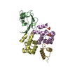 6ghdS S: Starting model for refinement |
|---|---|
| Similar structure data |

-Links
 PDBj
PDBj


- Assembly
| Deposited unit | 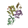
| ||||||||
|---|---|---|---|---|---|---|---|---|---|
| 1 |
| ||||||||
| Unit cell |
|
-Components
| #1: Protein | Mass: 12945.543 Da / Num. of mol.: 1 Source method: isolated from a genetically manipulated source Source: (gene. exp.) Homo sapiens (human) / Gene: LMNA, LMN1 / Production host:  | ||||||
|---|---|---|---|---|---|---|---|
| #2: Protein | Mass: 9713.026 Da / Num. of mol.: 2 Source method: isolated from a genetically manipulated source Source: (gene. exp.) Homo sapiens (human) / Production host: #3: Protein/peptide | | Mass: 5122.723 Da / Num. of mol.: 1 Source method: isolated from a genetically manipulated source Source: (gene. exp.) Homo sapiens (human) / Production host: #4: Chemical |   Mass: 96.063 Da / Num. of mol.: 3 / Source method: obtained synthetically / Formula: SO4 / Feature type: SUBJECT OF INVESTIGATION Mass: 96.063 Da / Num. of mol.: 3 / Source method: obtained synthetically / Formula: SO4 / Feature type: SUBJECT OF INVESTIGATION#5: Water | ChemComp-HOH / |  Mass: 18.015 Da / Num. of mol.: 83 / Source method: isolated from a natural source / Formula: H2O Mass: 18.015 Da / Num. of mol.: 83 / Source method: isolated from a natural source / Formula: H2O |
-Experimental details
-Experiment
| Experiment | Method: X-RAY DIFFRACTION / Number of used crystals: 2 |
|---|
- Sample preparation
| Crystal | Density Matthews: 2.41 Å3/Da / Density % sol: 49.05 % |
|---|---|
| Crystal grow | Temperature: 277 K / Method: vapor diffusion, hanging drop / Details: 18% PEG 3350, 100 mM Tris Bis pH 5.5, 0.1 M NH4SO4 |
-Data collection
| Diffraction | Mean temperature: 293 K / Serial crystal experiment: N |
|---|---|
| Diffraction source | Source: SYNCHROTRON / Site: SOLEIL / Beamline: PROXIMA 1 / Wavelength: 0.98 Å |
| Detector | Type: DECTRIS PILATUS3 S 6M / Detector: PIXEL / Date: Jun 10, 2017 |
| Radiation | Protocol: SINGLE WAVELENGTH / Monochromatic (M) / Laue (L): M / Scattering type: x-ray |
| Radiation wavelength | Wavelength: 0.98 Å / Relative weight: 1 |
| Reflection | Resolution: 2.26→40.74 Å / Num. obs: 16448 / % possible obs: 98.04 % / Redundancy: 4.5 % / Biso Wilson estimate: 55.09 Å2 / CC1/2: 0.997 / Rmerge(I) obs: 0.09554 / Rpim(I) all: 0.04968 / Rrim(I) all: 0.1061 / Net I/σ(I): 11.74 |
| Reflection shell | Resolution: 2.26→2.34 Å / Num. unique obs: 16448 |
- Processing
| Software |
| ||||||||||||||||||||||||||||||||||||||||||||||||||||||||||||||||||||||||||||||||||||||||||||||||||||||||||||
|---|---|---|---|---|---|---|---|---|---|---|---|---|---|---|---|---|---|---|---|---|---|---|---|---|---|---|---|---|---|---|---|---|---|---|---|---|---|---|---|---|---|---|---|---|---|---|---|---|---|---|---|---|---|---|---|---|---|---|---|---|---|---|---|---|---|---|---|---|---|---|---|---|---|---|---|---|---|---|---|---|---|---|---|---|---|---|---|---|---|---|---|---|---|---|---|---|---|---|---|---|---|---|---|---|---|---|---|---|---|
| Refinement | Method to determine structure: MOLECULAR REPLACEMENT Starting model: 6GHD Resolution: 2.26→40 Å / Cor.coef. Fo:Fc: 0.94 / Cor.coef. Fo:Fc free: 0.897 / SU R Cruickshank DPI: 0.314 / Cross valid method: THROUGHOUT / σ(F): 0 / SU R Blow DPI: 0.302 / SU Rfree Blow DPI: 0.22 / SU Rfree Cruickshank DPI: 0.226
| ||||||||||||||||||||||||||||||||||||||||||||||||||||||||||||||||||||||||||||||||||||||||||||||||||||||||||||
| Displacement parameters | Biso max: 159.53 Å2 / Biso mean: 47.15 Å2 / Biso min: 26.23 Å2
| ||||||||||||||||||||||||||||||||||||||||||||||||||||||||||||||||||||||||||||||||||||||||||||||||||||||||||||
| Refine analyze | Luzzati coordinate error obs: 0.29 Å | ||||||||||||||||||||||||||||||||||||||||||||||||||||||||||||||||||||||||||||||||||||||||||||||||||||||||||||
| Refinement step | Cycle: final / Resolution: 2.26→40 Å
| ||||||||||||||||||||||||||||||||||||||||||||||||||||||||||||||||||||||||||||||||||||||||||||||||||||||||||||
| Refine LS restraints |
| ||||||||||||||||||||||||||||||||||||||||||||||||||||||||||||||||||||||||||||||||||||||||||||||||||||||||||||
| LS refinement shell | Resolution: 2.26→2.42 Å / Rfactor Rfree error: 0 / Total num. of bins used: 8
|
