 Movie
Movie Controller

[English] 日本語
 Yorodumi
- PDB-2q16: Structure of the E. coli inosine triphosphate pyrophosphatase Rgd... -
+ Open data

- Basic information
| Entry | Database: PDB / ID: 2q16 | ||||||
|---|---|---|---|---|---|---|---|
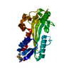
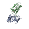
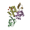
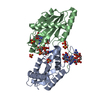
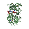
| Title | Structure of the E. coli inosine triphosphate pyrophosphatase RgdB in complex with ITP | ||||||
 Components Components | HAM1 protein homolog | ||||||
 Keywords Keywords | HYDROLASE / ITP pyrophosphatase x-ray structure enzyme mechanism substrate | ||||||
| Function / homology |  Function and homology information Function and homology informationXTP/dITP diphosphatase / purine nucleoside triphosphate catabolic process / ITP diphosphatase activity / XTP diphosphatase activity / dITP diphosphatase activity / nucleoside triphosphate catabolic process / nucleoside triphosphate diphosphatase activity / ribonucleoside triphosphate phosphatase activity / magnesium ion binding / protein homodimerization activity / cytosol Similarity search - Function | ||||||
| Biological species |   Escherichia coli (E. coli) Escherichia coli (E. coli) | ||||||
| Method |  X-RAY DIFFRACTION / SYNCHROTRON / SAD / Resolution: 1.95 Å X-RAY DIFFRACTION / SYNCHROTRON / SAD / Resolution: 1.95 Å | ||||||
 Authors Authors | Singer, A.U. / Lam, R. / Proudfoot, M. / Skarina, T. / Savchenko, A. / Yakunin, A.F. | ||||||
 Citation Citation | Journal: J.Mol.Biol. / Year: 2007 Title: Molecular basis of the antimutagenic activity of the house-cleaning inosine triphosphate pyrophosphatase RdgB from Escherichia coli. Authors: Savchenko, A. / Proudfoot, M. / Skarina, T. / Singer, A. / Litvinova, O. / Sanishvili, R. / Brown, G. / Chirgadze, N. / Yakunin, A.F. | ||||||
| History |
| ||||||
| Remark 999 | sequence These residues belong to a TEV cleavage site. |
- Structure visualization
| Structure viewer | Molecule: MolmilJmol/JSmol |
|---|
- Downloads & links
-Download
| PDBx/mmCIF format | 2q16.cif.gz | 100.3 KB | Display | PDBx/mmCIF format |
|---|---|---|---|---|
| PDB format | pdb2q16.ent.gz | 75 KB | Display | PDB format |
| PDBx/mmJSON format | 2q16.json.gz | Tree view | PDBx/mmJSON format | |
| Others |  Other downloads Other downloads |
-Validation report
| Arichive directory | https://data.pdbj.org/pub/pdb/validation_reports/q1/2q16ftp://data.pdbj.org/pub/pdb/validation_reports/q1/2q16 | HTTPS FTP |
|---|
-Related structure data



-Links
 PDBj
PDBj- Assembly
| Deposited unit | 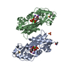
| ||||||||
|---|---|---|---|---|---|---|---|---|---|
| 1 |
| ||||||||
| Unit cell |
|
-Components
-Protein , 1 types, 2 molecules AB
| #1: Protein | Mass: 23475.980 Da / Num. of mol.: 2 / Mutation: D69A Source method: isolated from a genetically manipulated source Source: (gene. exp.) Escherichia coli (E. coli) / Strain: K12 / Gene: yggV / Plasmid: pET15-b-based p11 / Production host: Escherichia coli (E. coli) / Strain (production host): BL21(DE3) Codon Plus RPReferences: UniProt: P52061, nucleoside-triphosphate diphosphatase |
|---|
-Non-polymers , 5 types, 420 molecules 


| #2: Chemical |  Mass: 40.078 Da / Num. of mol.: 2 / Source method: obtained synthetically / Formula: Ca Mass: 40.078 Da / Num. of mol.: 2 / Source method: obtained synthetically / Formula: Ca#3: Chemical | ChemComp-NA /  Mass: 22.990 Da / Num. of mol.: 5 / Source method: obtained synthetically / Formula: Na Mass: 22.990 Da / Num. of mol.: 5 / Source method: obtained synthetically / Formula: Na#4: Chemical | ChemComp-SO4 / | 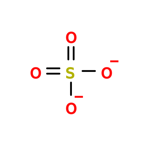 Mass: 96.063 Da / Num. of mol.: 1 / Source method: obtained synthetically / Formula: SO4 Mass: 96.063 Da / Num. of mol.: 1 / Source method: obtained synthetically / Formula: SO4#5: Chemical | 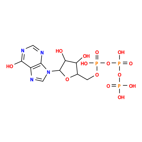 Mass: 508.166 Da / Num. of mol.: 2 / Source method: obtained synthetically / Formula: C10H15N4O14P3 Mass: 508.166 Da / Num. of mol.: 2 / Source method: obtained synthetically / Formula: C10H15N4O14P3#6: Water | ChemComp-HOH / | Mass: 18.015 Da / Num. of mol.: 410 / Source method: isolated from a natural source / Formula: H2O |
|---|
-Details
| Has protein modification | N |
|---|
-Experimental details
-Experiment
| Experiment | Method: X-RAY DIFFRACTION / Number of used crystals: 1 |
|---|
- Sample preparation
| Crystal | Density Matthews: 2.58 Å3/Da / Density % sol: 52.29 % |
|---|---|
| Crystal grow | Temperature: 298 K / Method: vapor diffusion, hanging drop / pH: 4.6 Details: 25% PEG 4K, 200 mM Ammonium Sulfate, 100 mM Sodium Acetate, 2% iso-Propanol, 20 mM Calcium Chloride, 10mM ITP, pH 4.6, VAPOR DIFFUSION, HANGING DROP, temperature 298K |
-Data collection
| Diffraction | Mean temperature: 100 K |
|---|---|
| Diffraction source | Source: SYNCHROTRON / Site: APS  / Beamline: 19-ID / Wavelength: 0.97942 Å / Beamline: 19-ID / Wavelength: 0.97942 Å |
| Detector | Type: ADSC QUANTUM 315 / Detector: CCD / Date: Dec 18, 2006 / Details: mirrors |
| Radiation | Monochromator: YALE MIRRORS / Protocol: SINGLE WAVELENGTH / Monochromatic (M) / Laue (L): M / Scattering type: x-ray |
| Radiation wavelength | Wavelength: 0.97942 Å / Relative weight: 1 |
| Reflection | Resolution: 1.95→500 Å / Num. all: 36421 / Num. obs: 36385 / % possible obs: 99.96 % / Redundancy: 19.4 % / Biso Wilson estimate: 24.8 Å2 / Rmerge(I) obs: 0.089 / Net I/σ(I): 39.9 |
| Reflection shell | Resolution: 1.95→2 Å / Redundancy: 17.7 % / Rmerge(I) obs: 0.551 / Mean I/σ(I) obs: 5.5 / Num. unique all: 2359 / % possible all: 100 |
- Processing
| Software |
| |||||||||||||||||||||||||||||||||||||||||||||||||||||||||||||||||||||||||||||||||||||||||||||||
|---|---|---|---|---|---|---|---|---|---|---|---|---|---|---|---|---|---|---|---|---|---|---|---|---|---|---|---|---|---|---|---|---|---|---|---|---|---|---|---|---|---|---|---|---|---|---|---|---|---|---|---|---|---|---|---|---|---|---|---|---|---|---|---|---|---|---|---|---|---|---|---|---|---|---|---|---|---|---|---|---|---|---|---|---|---|---|---|---|---|---|---|---|---|---|---|---|
| Refinement | Method to determine structure: SAD / Resolution: 1.95→26.98 Å / Cor.coef. Fo:Fc: 0.959 / Cor.coef. Fo:Fc free: 0.941 / SU B: 4.965 / SU ML: 0.081 / TLS residual ADP flag: LIKELY RESIDUAL / Isotropic thermal model: isotropic / Cross valid method: THROUGHOUT / ESU R: 0.137 / ESU R Free: 0.13 / Stereochemistry target values: MAXIMUM LIKELIHOOD
| |||||||||||||||||||||||||||||||||||||||||||||||||||||||||||||||||||||||||||||||||||||||||||||||
| Solvent computation | Ion probe radii: 0.8 Å / Shrinkage radii: 0.8 Å / VDW probe radii: 1.2 Å / Solvent model: MASK | |||||||||||||||||||||||||||||||||||||||||||||||||||||||||||||||||||||||||||||||||||||||||||||||
| Displacement parameters | Biso mean: 29.725 Å2
| |||||||||||||||||||||||||||||||||||||||||||||||||||||||||||||||||||||||||||||||||||||||||||||||
| Refinement step | Cycle: LAST / Resolution: 1.95→26.98 Å
| |||||||||||||||||||||||||||||||||||||||||||||||||||||||||||||||||||||||||||||||||||||||||||||||
| Refine LS restraints |
| |||||||||||||||||||||||||||||||||||||||||||||||||||||||||||||||||||||||||||||||||||||||||||||||
| LS refinement shell | Resolution: 1.95→2.001 Å / Total num. of bins used: 20
| |||||||||||||||||||||||||||||||||||||||||||||||||||||||||||||||||||||||||||||||||||||||||||||||
| Refinement TLS params. | Method: refined / Refine-ID: X-RAY DIFFRACTION
| |||||||||||||||||||||||||||||||||||||||||||||||||||||||||||||||||||||||||||||||||||||||||||||||
| Refinement TLS group |
|
