 Movie
Movie Controller

[English] 日本語
 Yorodumi
- PDB-6rnd: Liquid Application Method for time-resolved Analyses (LAMA) by se... -
+ Open data

- Basic information
| Entry | Database: PDB / ID: 6rnd | ||||||
|---|---|---|---|---|---|---|---|
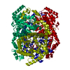
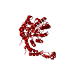
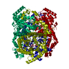
| Title | Liquid Application Method for time-resolved Analyses (LAMA) by serial synchrotron crystallography, Xylose Isomerase 15 ms timepoint | ||||||
 Components Components | Xylose isomerase | ||||||
 Keywords Keywords | ISOMERASE / Complex / Diffusion / Dynamics | ||||||
| Function / homology |  Function and homology information Function and homology informationxylose isomerase / xylose isomerase activity / D-xylose metabolic process / magnesium ion binding / identical protein binding / cytoplasm Similarity search - Function | ||||||
| Biological species |  Streptomyces rubiginosus (bacteria) Streptomyces rubiginosus (bacteria) | ||||||
| Method |  X-RAY DIFFRACTION / SYNCHROTRON / MOLECULAR REPLACEMENT / Resolution: 1.7 Å X-RAY DIFFRACTION / SYNCHROTRON / MOLECULAR REPLACEMENT / Resolution: 1.7 Å | ||||||
 Authors Authors | Mehrabi, P. / Schulz, E.C. / Miller, R.J.D. | ||||||
 Citation Citation | Journal: Nat.Methods / Year: 2019 Title: Liquid application method for time-resolved analyses by serial synchrotron crystallography. Authors: Mehrabi, P. / Schulz, E.C. / Agthe, M. / Horrell, S. / Bourenkov, G. / von Stetten, D. / Leimkohl, J.P. / Schikora, H. / Schneider, T.R. / Pearson, A.R. / Tellkamp, F. / Miller, R.J.D. | ||||||
| History |
|
- Structure visualization
| Structure viewer | Molecule: MolmilJmol/JSmol |
|---|
- Downloads & links
-Download
| PDBx/mmCIF format | 6rnd.cif.gz | 96.3 KB | Display | PDBx/mmCIF format |
|---|---|---|---|---|
| PDB format | pdb6rnd.ent.gz | 71.3 KB | Display | PDB format |
| PDBx/mmJSON format | 6rnd.json.gz | Tree view | PDBx/mmJSON format | |
| Others |  Other downloads Other downloads |
-Validation report
| Summary document | 6rnd_validation.pdf.gz | 1.6 MB | Display | wwPDB validaton report |
|---|---|---|---|---|
| Full document | 6rnd_full_validation.pdf.gz | 1.6 MB | Display | |
| Data in XML | 6rnd_validation.xml.gz | 17.9 KB | Display | |
| Data in CIF | 6rnd_validation.cif.gz | 26.6 KB | Display | |
| Arichive directory | https://data.pdbj.org/pub/pdb/validation_reports/rn/6rndftp://data.pdbj.org/pub/pdb/validation_reports/rn/6rnd | HTTPS FTP |
-Related structure data
| Related structure data |  6qnbC  6qncC  6qndC  6qnhC  6qniC  6qnjC  6rnbC  6rncC  6rnfC  3kbnS S: Starting model for refinement C: citing same article ( |
|---|---|
| Similar structure data |

-Links
 PDBj
PDBj


- Assembly
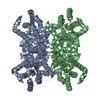
| Deposited unit | 
| |||||||||
|---|---|---|---|---|---|---|---|---|---|---|
| 1 | 
| |||||||||
| Unit cell |
| |||||||||
| Components on special symmetry positions |
|
-Components
| #1: Protein | Mass: 43283.297 Da / Num. of mol.: 1 Source method: isolated from a genetically manipulated source Source: (gene. exp.) Streptomyces rubiginosus (bacteria) / Gene: xylA / Production host: Streptomyces rubiginosus (bacteria) / References: UniProt: P24300, xylose isomerase | ||||
|---|---|---|---|---|---|
| #2: Chemical |   Mass: 24.305 Da / Num. of mol.: 2 / Source method: obtained synthetically / Formula: Mg / Feature type: SUBJECT OF INVESTIGATION Mass: 24.305 Da / Num. of mol.: 2 / Source method: obtained synthetically / Formula: Mg / Feature type: SUBJECT OF INVESTIGATION#3: Sugar | ChemComp-GLC / |   Type: D-saccharide, alpha linking / Mass: 180.156 Da / Num. of mol.: 1 Type: D-saccharide, alpha linking / Mass: 180.156 Da / Num. of mol.: 1Source method: isolated from a genetically manipulated source Formula: C6H12O6 / Feature type: SUBJECT OF INVESTIGATION #4: Water | ChemComp-HOH / |  Mass: 18.015 Da / Num. of mol.: 243 / Source method: isolated from a natural source / Formula: H2O Mass: 18.015 Da / Num. of mol.: 243 / Source method: isolated from a natural source / Formula: H2O |
-Experimental details
-Experiment
| Experiment | Method: X-RAY DIFFRACTION / Number of used crystals: 1 |
|---|
- Sample preparation
| Crystal | Density Matthews: 2.38 Å3/Da / Density % sol: 48.29 % |
|---|---|
| Crystal grow | Temperature: 293 K / Method: batch mode Details: 35% (w/v) PEG3350, 200 mM lithium sulfate and 10 mM Hepes/NaOH pH 7.5 |
-Data collection
| Diffraction | Mean temperature: 293 K / Serial crystal experiment: N |
|---|---|
| Diffraction source | Source: SYNCHROTRON / Site: PETRA III, EMBL c/o DESY  / Beamline: P14 (MX2) / Wavelength: 0.98 Å / Beamline: P14 (MX2) / Wavelength: 0.98 Å |
| Detector | Type: DECTRIS PILATUS 2M / Detector: PIXEL / Date: May 1, 2019 |
| Radiation | Protocol: SINGLE WAVELENGTH / Monochromatic (M) / Laue (L): M / Scattering type: x-ray |
| Radiation wavelength | Wavelength: 0.98 Å / Relative weight: 1 |
| Reflection | Resolution: 1.699→68.526 Å / Num. obs: 45700 / % possible obs: 99.99 % / Redundancy: 492.9 % / Net I/σ(I): 5.36 |
| Reflection shell | Resolution: 1.699→1.737 Å / Num. unique obs: 4499 |
- Processing
| Software |
| |||||||||||||||||||||||||||||||||||||||||||||||||||||||||||||||||||||||||||||||||||||||||||||||||||||||||||||||||||||||
|---|---|---|---|---|---|---|---|---|---|---|---|---|---|---|---|---|---|---|---|---|---|---|---|---|---|---|---|---|---|---|---|---|---|---|---|---|---|---|---|---|---|---|---|---|---|---|---|---|---|---|---|---|---|---|---|---|---|---|---|---|---|---|---|---|---|---|---|---|---|---|---|---|---|---|---|---|---|---|---|---|---|---|---|---|---|---|---|---|---|---|---|---|---|---|---|---|---|---|---|---|---|---|---|---|---|---|---|---|---|---|---|---|---|---|---|---|---|---|---|---|
| Refinement | Method to determine structure: MOLECULAR REPLACEMENT Starting model: 3KBN Resolution: 1.7→68.526 Å / SU ML: 0.2 / Cross valid method: FREE R-VALUE / σ(F): 1.34 / Phase error: 20.13
| |||||||||||||||||||||||||||||||||||||||||||||||||||||||||||||||||||||||||||||||||||||||||||||||||||||||||||||||||||||||
| Solvent computation | Shrinkage radii: 0.9 Å / VDW probe radii: 1.11 Å | |||||||||||||||||||||||||||||||||||||||||||||||||||||||||||||||||||||||||||||||||||||||||||||||||||||||||||||||||||||||
| Refinement step | Cycle: LAST / Resolution: 1.7→68.526 Å
| |||||||||||||||||||||||||||||||||||||||||||||||||||||||||||||||||||||||||||||||||||||||||||||||||||||||||||||||||||||||
| Refine LS restraints |
| |||||||||||||||||||||||||||||||||||||||||||||||||||||||||||||||||||||||||||||||||||||||||||||||||||||||||||||||||||||||
| LS refinement shell |
|
