 Movie
Movie Controller

[English] 日本語
 Yorodumi
- PDB-6rmw: Structure of N-terminal truncated IMP bound Plasmodium falciparum... -
+ Open data

- Basic information
| Entry | Database: PDB / ID: 6rmw | ||||||
|---|---|---|---|---|---|---|---|
| Title | Structure of N-terminal truncated IMP bound Plasmodium falciparum IMP-nucleotidase | ||||||
 Components Components | IMP-specific 5'-nucleotidase, putative | ||||||
 Keywords Keywords | HYDROLASE / nucleotidase / activator / complex | ||||||
| Function / homology |  Function and homology information Function and homology informationnicotinamide riboside biosynthetic process / nicotinic acid riboside biosynthetic process / inosine salvage / IMP-specific 5'-nucleotidase / IMP catabolic process / 5'-nucleotidase activity / magnesium ion binding / ATP binding / cytoplasm Similarity search - Function | ||||||
| Biological species |  | ||||||
| Method |  X-RAY DIFFRACTION / SYNCHROTRON / MOLECULAR REPLACEMENT / Resolution: 3.5 Å X-RAY DIFFRACTION / SYNCHROTRON / MOLECULAR REPLACEMENT / Resolution: 3.5 Å | ||||||
 Authors Authors | Carrique, L. / Ballut, L. / Violot, S. / Aghajari, N. | ||||||
| Funding support |  France, 1items France, 1items
| ||||||
 Citation Citation | Journal: Nat Commun / Year: 2020 Title: Structure and catalytic regulation of Plasmodium falciparum IMP specific nucleotidase. Authors: Carrique, L. / Ballut, L. / Shukla, A. / Varma, N. / Ravi, R. / Violot, S. / Srinivasan, B. / Ganeshappa, U.T. / Kulkarni, S. / Balaram, H. / Aghajari, N. | ||||||
| History |
|
- Structure visualization
| Structure viewer | Molecule: MolmilJmol/JSmol |
|---|
- Downloads & links
-Download
| PDBx/mmCIF format | 6rmw.cif.gz | 598.2 KB | Display | PDBx/mmCIF format |
|---|---|---|---|---|
| PDB format | pdb6rmw.ent.gz | 481.9 KB | Display | PDB format |
| PDBx/mmJSON format | 6rmw.json.gz | Tree view | PDBx/mmJSON format | |
| Others |  Other downloads Other downloads |
-Validation report
| Arichive directory | https://data.pdbj.org/pub/pdb/validation_reports/rm/6rmwftp://data.pdbj.org/pub/pdb/validation_reports/rm/6rmw | HTTPS FTP |
|---|
-Related structure data
| Related structure data |  6rmdSC 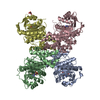 6rmeC 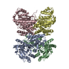 6rmoC  6rn1C 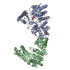 6rnhC C: citing same article ( S: Starting model for refinement |
|---|---|
| Similar structure data |

-Links
 PDBj
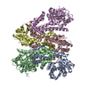
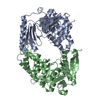
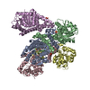
PDBj- Assembly
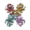
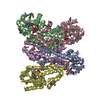
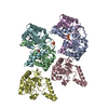
| Deposited unit | 
| ||||||||
|---|---|---|---|---|---|---|---|---|---|
| 1 | 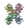
| ||||||||
| 2 | 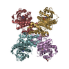
| ||||||||
| Unit cell |
|
-Components
| #1: Protein | Mass: 48496.832 Da / Num. of mol.: 8 / Mutation: D172N Source method: isolated from a genetically manipulated source Details: First 30 residues are truncated in the construction which also contains the mutation D172N Source: (gene. exp.)  #2: Chemical | ChemComp-IMP /   Mass: 348.206 Da / Num. of mol.: 8 / Source method: obtained synthetically / Formula: C10H13N4O8P Mass: 348.206 Da / Num. of mol.: 8 / Source method: obtained synthetically / Formula: C10H13N4O8P#3: Chemical | ChemComp-MG /   Mass: 24.305 Da / Num. of mol.: 8 / Source method: obtained synthetically / Formula: Mg Mass: 24.305 Da / Num. of mol.: 8 / Source method: obtained synthetically / Formula: Mg#4: Chemical |   Mass: 92.094 Da / Num. of mol.: 2 / Source method: obtained synthetically / Formula: C3H8O3 Mass: 92.094 Da / Num. of mol.: 2 / Source method: obtained synthetically / Formula: C3H8O3Has ligand of interest | N | Has protein modification | N | |
|---|
-Experimental details
-Experiment
| Experiment | Method: X-RAY DIFFRACTION / Number of used crystals: 1 |
|---|
- Sample preparation
| Crystal | Density Matthews: 3 Å3/Da / Density % sol: 58.97 % |
|---|---|
| Crystal grow | Temperature: 292 K / Method: vapor diffusion, sitting drop Details: 0.1 M HEPES pH 7.5, 0.2 M calcium acetate, 10% (w/v) PEG 8000 by co-crystallizing the protein with 5 mM of IMP |
-Data collection
| Diffraction | Mean temperature: 100 K / Serial crystal experiment: N |
|---|---|
| Diffraction source | Source: SYNCHROTRON / Site: ESRF / Beamline: ID30B / Wavelength: 1 Å |
| Detector | Type: DECTRIS PILATUS3 6M / Detector: PIXEL / Date: Dec 9, 2016 |
| Radiation | Protocol: SINGLE WAVELENGTH / Monochromatic (M) / Laue (L): M / Scattering type: x-ray |
| Radiation wavelength | Wavelength: 1 Å / Relative weight: 1 |
| Reflection | Resolution: 3.5→49.7 Å / Num. obs: 57015 / % possible obs: 99.14 % / Redundancy: 6.7 % / CC1/2: 0.76 / Net I/σ(I): 4.2 |
| Reflection shell | Resolution: 3.5→4.3 Å / Num. unique obs: 26211 / CC1/2: 0.767 |
- Processing
| Software |
| ||||||||||||||||||||||||||||||||||||||||||||||||||||||||||||||||||||||||||||||||||||||||||||||||||||||||||||||||||||||||||||||||||||||||||||
|---|---|---|---|---|---|---|---|---|---|---|---|---|---|---|---|---|---|---|---|---|---|---|---|---|---|---|---|---|---|---|---|---|---|---|---|---|---|---|---|---|---|---|---|---|---|---|---|---|---|---|---|---|---|---|---|---|---|---|---|---|---|---|---|---|---|---|---|---|---|---|---|---|---|---|---|---|---|---|---|---|---|---|---|---|---|---|---|---|---|---|---|---|---|---|---|---|---|---|---|---|---|---|---|---|---|---|---|---|---|---|---|---|---|---|---|---|---|---|---|---|---|---|---|---|---|---|---|---|---|---|---|---|---|---|---|---|---|---|---|---|---|
| Refinement | Method to determine structure: MOLECULAR REPLACEMENT Starting model: 6RMD Resolution: 3.5→48.073 Å / SU ML: 0.77 / Cross valid method: FREE R-VALUE / σ(F): 1.33 / Phase error: 34.95 / Stereochemistry target values: ML
| ||||||||||||||||||||||||||||||||||||||||||||||||||||||||||||||||||||||||||||||||||||||||||||||||||||||||||||||||||||||||||||||||||||||||||||
| Solvent computation | Shrinkage radii: 0.9 Å / VDW probe radii: 1.11 Å / Solvent model: FLAT BULK SOLVENT MODEL | ||||||||||||||||||||||||||||||||||||||||||||||||||||||||||||||||||||||||||||||||||||||||||||||||||||||||||||||||||||||||||||||||||||||||||||
| Refinement step | Cycle: LAST / Resolution: 3.5→48.073 Å
| ||||||||||||||||||||||||||||||||||||||||||||||||||||||||||||||||||||||||||||||||||||||||||||||||||||||||||||||||||||||||||||||||||||||||||||
| Refine LS restraints |
| ||||||||||||||||||||||||||||||||||||||||||||||||||||||||||||||||||||||||||||||||||||||||||||||||||||||||||||||||||||||||||||||||||||||||||||
| LS refinement shell |
|
