[English] 日本語
Yorodumi
- PDB-6rlz: Crystal Structure of 14-3-3zeta in complex with a macrocyclic 8-m... -

+
Open data


ID or keywords:

Loading...

-
Basic information

Entry
Database: PDB / ID: 6rlz
Title
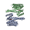
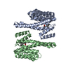
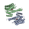
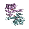
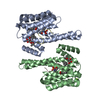
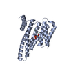
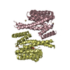
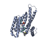
Crystal Structure of 14-3-3zeta in complex with a macrocyclic 8-mer peptide derived from ExoS
Components
  • 14-3-3 protein zeta/delta
  • EtMe
KeywordsPROTEIN BINDING / protein-protein-interaction macrocycle peptide
Function / homology
Function and homology information


Golgi reassembly / regulation of synapse maturation / NOTCH4 Activation and Transmission of Signal to the Nucleus / establishment of Golgi localization / Rap1 signalling / negative regulation of protein localization to nucleus / KSRP (KHSRP) binds and destabilizes mRNA / GP1b-IX-V activation signalling / Regulation of localization of FOXO transcription factors / Interleukin-3, Interleukin-5 and GM-CSF signaling ...Golgi reassembly / regulation of synapse maturation / NOTCH4 Activation and Transmission of Signal to the Nucleus / establishment of Golgi localization / Rap1 signalling / negative regulation of protein localization to nucleus / KSRP (KHSRP) binds and destabilizes mRNA / GP1b-IX-V activation signalling / Regulation of localization of FOXO transcription factors / Interleukin-3, Interleukin-5 and GM-CSF signaling / phosphoserine residue binding / Activation of BAD and translocation to mitochondria / cellular response to glucose starvation / SARS-CoV-2 targets host intracellular signalling and regulatory pathways / Chk1/Chk2(Cds1) mediated inactivation of Cyclin B:Cdk1 complex / SARS-CoV-1 targets host intracellular signalling and regulatory pathways / RHO GTPases activate PKNs / negative regulation of TORC1 signaling / negative regulation of innate immune response / regulation of ERK1 and ERK2 cascade / protein sequestering activity / Translocation of SLC2A4 (GLUT4) to the plasma membrane / Deactivation of the beta-catenin transactivating complex / TP53 Regulates Metabolic Genes / Negative regulation of NOTCH4 signaling / melanosome / blood microparticle / DNA-binding transcription factor binding / vesicle / transmembrane transporter binding / cadherin binding / protein phosphorylation / focal adhesion / glutamatergic synapse / ubiquitin protein ligase binding / negative regulation of apoptotic process / protein kinase binding / negative regulation of transcription by RNA polymerase II / signal transduction / extracellular space / RNA binding / extracellular exosome / nucleoplasm / identical protein binding / nucleus / cytosol / cytoplasm
Similarity search - Function
14-3-3 domain / Delta-Endotoxin; domain 1 / 14-3-3 proteins signature 2. / 14-3-3 protein, conserved site / 14-3-3 proteins signature 1. / 14-3-3 protein / 14-3-3 homologues / 14-3-3 domain / 14-3-3 domain superfamily / 14-3-3 protein ...14-3-3 domain / Delta-Endotoxin; domain 1 / 14-3-3 proteins signature 2. / 14-3-3 protein, conserved site / 14-3-3 proteins signature 1. / 14-3-3 protein / 14-3-3 homologues / 14-3-3 domain / 14-3-3 domain superfamily / 14-3-3 protein / Up-down Bundle / Mainly Alpha
Similarity search - Domain/homology
14-3-3 protein zeta/delta
Similarity search - Component
Biological speciesHomo sapiens (human)
Pseudomonas aeruginosa (bacteria)
MethodX-RAY DIFFRACTION / SYNCHROTRON / MOLECULAR REPLACEMENT / Resolution: 3.7 Å
AuthorsWallraven, K. / Grossmann, T.N. / Hennig, S.
Funding support1items
OrganizationGrant numberCountry
European Research Council
CitationJournal: Chem Sci / Year: 2020
Title: Adapting free energy perturbation simulations for large macrocyclic ligands: how to dissect contributions from direct binding and free ligand flexibility.
Authors: Wallraven, K. / Holmelin, F.L. / Glas, A. / Hennig, S. / Frolov, A.I. / Grossmann, T.N.
History
DepositionMay 3, 2019Deposition site: PDBE / Processing site: PDBE
Revision 1.0Mar 18, 2020Provider: repository / Type: Initial release
Revision 1.1Mar 25, 2020Group: Database references / Category: citation / citation_author
Item: _citation.journal_id_ISSN / _citation.pdbx_database_id_DOI ..._citation.journal_id_ISSN / _citation.pdbx_database_id_DOI / _citation.pdbx_database_id_PubMed / _citation.title / _citation_author.identifier_ORCID / _citation_author.name

-
Structure visualization

Structure viewerMolecule:
MolmilJmol/JSmol

Downloads & links

-
Assembly

Deposited unit
A: 14-3-3 protein zeta/delta
B: 14-3-3 protein zeta/delta
C: EtMe
D: EtMe


Theoretical massNumber of molelcules
Total (without water)54,4604
Polymers54,4604
Non-polymers00
Water50428
1


  • Idetical with deposited unit
  • defined by author&software
TypeNameSymmetry operationNumber
identity operation1_555x,y,z1
Buried area3880 Å2
ΔGint-19 kcal/mol
Surface area21160 Å2
MethodPISA
Unit cell
Length a, b, c (Å)98.800, 98.800, 94.800
Angle α, β, γ (deg.)90.00, 90.00, 120.00
Int Tables number172
Space group name H-MP64

-
Components

#1: Protein 14-3-3 protein zeta/delta / Protein kinase C inhibitor protein 1 / KCIP-1


Mass: 26316.764 Da / Num. of mol.: 2
Source method: isolated from a genetically manipulated source
Source: (gene. exp.) Homo sapiens (human) / Gene: YWHAZ / Plasmid: pProEx HTb / Production host: Escherichia coli BL21(DE3) (bacteria) / References: UniProt: P63104
#2: Protein/peptide EtMe


Mass: 913.111 Da / Num. of mol.: 2 / Source method: obtained synthetically / Source: (synth.) Pseudomonas aeruginosa (bacteria)
#3: Water ChemComp-HOH / water / Water


Mass: 18.015 Da / Num. of mol.: 28 / Source method: isolated from a natural source / Formula: H2O

-
Experimental details

-
Experiment

ExperimentMethod: X-RAY DIFFRACTION / Number of used crystals: 1

-
Sample preparation

CrystalDensity Matthews: 2.52 Å3/Da / Density % sol: 51.16 %
Crystal growTemperature: 277 K / Method: vapor diffusion, hanging drop / Details: 0.2 M Magnesium formate 20% PEG 3350 pH 7.5

-
Data collection

DiffractionMean temperature: 100 K / Serial crystal experiment: N
Diffraction sourceSource: SYNCHROTRON / Site: Diamond / Beamline: I24 / Wavelength: 0.9787 Å
DetectorType: DECTRIS PILATUS3 6M / Detector: PIXEL / Date: Dec 1, 2018
RadiationProtocol: SINGLE WAVELENGTH / Monochromatic (M) / Laue (L): M / Scattering type: x-ray
Radiation wavelengthWavelength: 0.9787 Å / Relative weight: 1
ReflectionResolution: 3.7→50 Å / Num. obs: 5633 / % possible obs: 99.1 % / Redundancy: 33 % / CC1/2: 0.992 / Net I/σ(I): 16.02
Reflection shellResolution: 3.7→10 Å / Num. unique obs: 186022 / CC1/2: 0.921

-
Processing

Software
NameVersionClassification
REFMAC5.8.0238refinement
XDSdata reduction
XDSdata scaling
PHASERphasing
RefinementMethod to determine structure: MOLECULAR REPLACEMENT / Resolution: 3.7→43.81 Å / Cor.coef. Fo:Fc: 0.952 / Cor.coef. Fo:Fc free: 0.908 / SU B: 95.852 / SU ML: 0.701 / Cross valid method: FREE R-VALUE / ESU R Free: 0.934
RfactorNum. reflection% reflectionSelection details
Rfree0.2922 564 10 %RANDOM
Rwork0.21599 ---
obs0.22336 5069 99.1 %-
Solvent computationIon probe radii: 0.8 Å / Shrinkage radii: 0.8 Å / VDW probe radii: 1.2 Å
Displacement parametersBiso mean: 120.233 Å2
Baniso -1Baniso -2Baniso -3
1--1.22 Å2-0.61 Å2-0 Å2
2---1.22 Å20 Å2
3---3.95 Å2
Refinement stepCycle: LAST / Resolution: 3.7→43.81 Å
ProteinNucleic acidLigandSolventTotal
Num. atoms3466 0 0 28 3494
Refine LS restraints
Refine-IDTypeDev idealDev ideal targetNumber
X-RAY DIFFRACTIONr_bond_refined_d0.0020.0123520
X-RAY DIFFRACTIONr_bond_other_d
X-RAY DIFFRACTIONr_angle_refined_deg0.8411.6394749
X-RAY DIFFRACTIONr_angle_other_deg
X-RAY DIFFRACTIONr_dihedral_angle_1_deg4.555457
X-RAY DIFFRACTIONr_dihedral_angle_2_deg37.38123.455165
X-RAY DIFFRACTIONr_dihedral_angle_3_deg21.80515554
X-RAY DIFFRACTIONr_dihedral_angle_4_deg14.9241518
X-RAY DIFFRACTIONr_chiral_restr0.0730.2486
X-RAY DIFFRACTIONr_gen_planes_refined0.0030.022622
X-RAY DIFFRACTIONr_gen_planes_other
X-RAY DIFFRACTIONr_nbd_refined
X-RAY DIFFRACTIONr_nbd_other
X-RAY DIFFRACTIONr_nbtor_refined
X-RAY DIFFRACTIONr_nbtor_other
X-RAY DIFFRACTIONr_xyhbond_nbd_refined
X-RAY DIFFRACTIONr_xyhbond_nbd_other
X-RAY DIFFRACTIONr_metal_ion_refined
X-RAY DIFFRACTIONr_metal_ion_other
X-RAY DIFFRACTIONr_symmetry_vdw_refined
X-RAY DIFFRACTIONr_symmetry_vdw_other
X-RAY DIFFRACTIONr_symmetry_hbond_refined
X-RAY DIFFRACTIONr_symmetry_hbond_other
X-RAY DIFFRACTIONr_symmetry_metal_ion_refined
X-RAY DIFFRACTIONr_symmetry_metal_ion_other
X-RAY DIFFRACTIONr_mcbond_it2.0738.781875
X-RAY DIFFRACTIONr_mcbond_other
X-RAY DIFFRACTIONr_mcangle_it3.52713.1152311
X-RAY DIFFRACTIONr_mcangle_other
X-RAY DIFFRACTIONr_scbond_it1.7338.7231645
X-RAY DIFFRACTIONr_scbond_other
X-RAY DIFFRACTIONr_scangle_it
X-RAY DIFFRACTIONr_scangle_other
X-RAY DIFFRACTIONr_long_range_B_refined8.3816006
X-RAY DIFFRACTIONr_long_range_B_other
X-RAY DIFFRACTIONr_rigid_bond_restr
X-RAY DIFFRACTIONr_sphericity_free
X-RAY DIFFRACTIONr_sphericity_bonded
LS refinement shellResolution: 3.7→3.796 Å / Total num. of bins used: 20
RfactorNum. reflection% reflection
Rfree0.474 42 -
Rwork0.38 375 -
obs--97.89 %
Refinement TLS params.

Method: refined / Refine-ID: X-RAY DIFFRACTION

IDL112)L122)L132)L222)L232)L332)S11 (Å °)S12 (Å °)S13 (Å °)S21 (Å °)S22 (Å °)S23 (Å °)S31 (Å °)S32 (Å °)S33 (Å °)T112)T122)T132)T222)T232)T332)Origin x (Å)Origin y (Å)Origin z (Å)
10.77950.1241.20692.7658-0.51522.4290.03810.11280.1399-0.4459-0.03390.78080.12490.5786-0.00420.08480.018-0.06860.414-0.13260.5268-38.950122.765341.4202
24.20310.95193.38481.5576-0.2676.69980.2584-0.1778-0.9951-0.1273-0.07740.13310.82640.5803-0.18090.30370.10260.07280.19590.18890.6222-24.3185-4.720360.4989
300000000000000-00.1488000.148800.1488000
Refinement TLS group
IDRefine-IDRefine TLS-IDAuth asym-IDAuth seq-ID
1X-RAY DIFFRACTION1A3 - 230
2X-RAY DIFFRACTION2B2 - 230

+
About Yorodumi

-
News

-
Feb 9, 2022. New format data for meta-information of EMDB entries

New format data for meta-information of EMDB entries

  • Version 3 of the EMDB header file is now the official format.
  • The previous official version 1.9 will be removed from the archive.

Related info.:EMDB header

External links:wwPDB to switch to version 3 of the EMDB data model

-
Aug 12, 2020. Covid-19 info

Covid-19 info

URL: https://pdbj.org/emnavi/covid19.php

New page: Covid-19 featured information page in EM Navigator.

Related info.:Covid-19 info / Mar 5, 2020. Novel coronavirus structure data

+
Mar 5, 2020. Novel coronavirus structure data

Novel coronavirus structure data

Related info.:Yorodumi Speices / Aug 12, 2020. Covid-19 info

External links:COVID-19 featured content - PDBj / Molecule of the Month (242):Coronavirus Proteases

+
Jan 31, 2019. EMDB accession codes are about to change! (news from PDBe EMDB page)

EMDB accession codes are about to change! (news from PDBe EMDB page)

  • The allocation of 4 digits for EMDB accession codes will soon come to an end. Whilst these codes will remain in use, new EMDB accession codes will include an additional digit and will expand incrementally as the available range of codes is exhausted. The current 4-digit format prefixed with “EMD-” (i.e. EMD-XXXX) will advance to a 5-digit format (i.e. EMD-XXXXX), and so on. It is currently estimated that the 4-digit codes will be depleted around Spring 2019, at which point the 5-digit format will come into force.
  • The EM Navigator/Yorodumi systems omit the EMD- prefix.

Related info.:Q: What is EMD? / ID/Accession-code notation in Yorodumi/EM Navigator

External links:EMDB Accession Codes are Changing Soon! / Contact to PDBj

+
Jul 12, 2017. Major update of PDB

Major update of PDB

  • wwPDB released updated PDB data conforming to the new PDBx/mmCIF dictionary.
  • This is a major update changing the version number from 4 to 5, and with Remediation, in which all the entries are updated.
  • In this update, many items about electron microscopy experimental information are reorganized (e.g. em_software).
  • Now, EM Navigator and Yorodumi are based on the updated data.

External links:wwPDB Remediation / Enriched Model Files Conforming to OneDep Data Standards Now Available in the PDB FTP Archive

-
Yorodumi

Thousand views of thousand structures

  • Yorodumi is a browser for structure data from EMDB, PDB, SASBDB, etc.
  • This page is also the successor to EM Navigator detail page, and also detail information page/front-end page for Omokage search.
  • The word "yorodu" (or yorozu) is an old Japanese word meaning "ten thousand". "mi" (miru) is to see.

Related info.:EMDB / PDB / SASBDB / Comparison of 3 databanks / Yorodumi Search / Aug 31, 2016. New EM Navigator & Yorodumi / Yorodumi Papers / Jmol/JSmol / Function and homology information / Changes in new EM Navigator and Yorodumi

Read more