 Movie
Movie Controller

[English] 日本語
 Yorodumi
- PDB-6r7t: Crystal Structure of human Melanoma-associated antigen B1 (MAGEB1... -
+ Open data

- Basic information
| Entry | Database: PDB / ID: 6r7t | ||||||
|---|---|---|---|---|---|---|---|
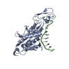
| Title | Crystal Structure of human Melanoma-associated antigen B1 (MAGEB1) in complex with nanobody | ||||||
 Components Components |
| ||||||
 Keywords Keywords | IMMUNE SYSTEM / Melanoma | ||||||
| Function / homology |  Function and homology information Function and homology information | ||||||
| Biological species |   Homo sapiens (human) Homo sapiens (human) | ||||||
| Method |  X-RAY DIFFRACTION / SYNCHROTRON / MOLECULAR REPLACEMENT / Resolution: 2.682 Å X-RAY DIFFRACTION / SYNCHROTRON / MOLECULAR REPLACEMENT / Resolution: 2.682 Å | ||||||
 Authors Authors | Ye, M. / Newman, J. / Pike, A.C.W. / Burgess-Brown, N. / Cooper, C.D.O. / Bountra, C. / Arrowsmith, C. / Edwards, A. / Gileadi, O. / von Delft, F. | ||||||
| Funding support |  United Kingdom, 1items United Kingdom, 1items
| ||||||
 Citation Citation | Journal: To Be Published Title: Crystal Structure of Melanoma-associated antigen B1 (MAGEB1) in complex with nanobody Authors: Ye, M. / Newman, J. / Pike, A.C.W. / Burgess-Brown, N. / Cooper, C.D.O. / Bountra, C. / Arrowsmith, C. / Edwards, A. / Gileadi, O. / von Delft, F. | ||||||
| History |
|
- Structure visualization
| Structure viewer | Molecule: MolmilJmol/JSmol |
|---|
- Downloads & links
-Download
| PDBx/mmCIF format | 6r7t.cif.gz | 76.7 KB | Display | PDBx/mmCIF format |
|---|---|---|---|---|
| PDB format | pdb6r7t.ent.gz | 55.3 KB | Display | PDB format |
| PDBx/mmJSON format | 6r7t.json.gz | Tree view | PDBx/mmJSON format | |
| Others |  Other downloads Other downloads |
-Validation report
| Arichive directory | https://data.pdbj.org/pub/pdb/validation_reports/r7/6r7tftp://data.pdbj.org/pub/pdb/validation_reports/r7/6r7t | HTTPS FTP |
|---|
-Related structure data
| Related structure data | 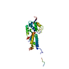 2wa0S S: Starting model for refinement |
|---|---|
| Similar structure data |


-Links
 PDBj
PDBj

- Assembly
| Deposited unit | 
| ||||||||
|---|---|---|---|---|---|---|---|---|---|
| 1 |
| ||||||||
| Unit cell |
|
-Components
| #1: Antibody | Mass: 13119.455 Da / Num. of mol.: 1 Source method: isolated from a genetically manipulated source Source: (gene. exp.) Production host:  |
|---|---|
| #2: Protein | Mass: 29203.293 Da / Num. of mol.: 1 Source method: isolated from a genetically manipulated source Source: (gene. exp.) Homo sapiens (human) / Gene: MAGEB1, MAGEL1, MAGEXPProduction host: References: UniProt: P43366 |
| #3: Water | ChemComp-HOH /  Mass: 18.015 Da / Num. of mol.: 43 / Source method: isolated from a natural source / Formula: H2O Mass: 18.015 Da / Num. of mol.: 43 / Source method: isolated from a natural source / Formula: H2O |
| Has protein modification | Y |
-Experimental details
-Experiment
| Experiment | Method: X-RAY DIFFRACTION / Number of used crystals: 1 |
|---|
- Sample preparation
| Crystal | Density Matthews: 2.36 Å3/Da / Density % sol: 47.98 % |
|---|---|
| Crystal grow | Temperature: 293 K / Method: vapor diffusion, sitting drop Details: 0.056M sodium phosphate monobasic - 1.344M potassium phosphate dibasic |
-Data collection
| Diffraction | Mean temperature: 100 K / Serial crystal experiment: N |
|---|---|
| Diffraction source | Source: SYNCHROTRON / Site: Diamond / Beamline: I24 / Wavelength: 0.9686 Å |
| Detector | Type: DECTRIS PILATUS3 6M / Detector: PIXEL / Date: Feb 3, 2019 |
| Radiation | Protocol: SINGLE WAVELENGTH / Monochromatic (M) / Laue (L): M / Scattering type: x-ray |
| Radiation wavelength | Wavelength: 0.9686 Å / Relative weight: 1 |
| Reflection | Resolution: 2.68→53.673 Å / Num. obs: 12641 / % possible obs: 100 % / Redundancy: 18.1 % / Rmerge(I) obs: 0.135 / Net I/σ(I): 12.1 |
| Reflection shell | Resolution: 2.68→2.81 Å / Num. unique obs: 1618 |
- Processing
| Software |
| |||||||||||||||||||||||||||||||||||
|---|---|---|---|---|---|---|---|---|---|---|---|---|---|---|---|---|---|---|---|---|---|---|---|---|---|---|---|---|---|---|---|---|---|---|---|---|
| Refinement | Method to determine structure: MOLECULAR REPLACEMENT Starting model: 2WA0 Resolution: 2.682→53.673 Å / SU ML: 0.45 / Cross valid method: FREE R-VALUE / σ(F): 1.35 / Phase error: 29.92
| |||||||||||||||||||||||||||||||||||
| Solvent computation | Shrinkage radii: 0.9 Å / VDW probe radii: 1.11 Å | |||||||||||||||||||||||||||||||||||
| Refinement step | Cycle: LAST / Resolution: 2.682→53.673 Å
| |||||||||||||||||||||||||||||||||||
| Refine LS restraints |
| |||||||||||||||||||||||||||||||||||
| LS refinement shell |
|
