 Movie
Movie Controller

[English] 日本語
 Yorodumi
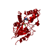
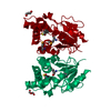
- PDB-6g2n: Crystal structure of human cytosolic 5'(3')-deoxyribonucleotidase... -
+ Open data

- Basic information
| Entry | Database: PDB / ID: 6g2n | ||||||||||||
|---|---|---|---|---|---|---|---|---|---|---|---|---|---|
| Title | Crystal structure of human cytosolic 5'(3')-deoxyribonucleotidase in complex with the inhibitor PB-PAU | ||||||||||||
 Components Components | 5'(3')-deoxyribonucleotidase, cytosolic type | ||||||||||||
 Keywords Keywords | HYDROLASE / 5'-nucleotidase / dephosphorylation / phosphorylation / HAD-like / mitochondria / HYDROLASE-HYDROLASE INHIBITOR complex | ||||||||||||
| Function / homology |  Function and homology information Function and homology informationpyrimidine nucleotide binding / pyrimidine deoxyribonucleotide catabolic process / dTMP catabolic process / dCMP catabolic process / nucleotidase activity / UMP catabolic process / dUMP catabolic process / : / Pyrimidine catabolism / dGMP catabolic process ...pyrimidine nucleotide binding / pyrimidine deoxyribonucleotide catabolic process / dTMP catabolic process / dCMP catabolic process / nucleotidase activity / UMP catabolic process / dUMP catabolic process / : / Pyrimidine catabolism / dGMP catabolic process / IMP catabolic process / allantoin metabolic process / Purine catabolism / Hydrolases; Acting on ester bonds; Phosphoric-monoester hydrolases / 5'-nucleotidase activity / dephosphorylation / mitochondrion / extracellular exosome / metal ion binding / identical protein binding / nucleus / cytoplasm / cytosol Similarity search - Function | ||||||||||||
| Biological species |  Homo sapiens (human) Homo sapiens (human) | ||||||||||||
| Method |  X-RAY DIFFRACTION / SYNCHROTRON / MOLECULAR REPLACEMENT / molecular replacement / Resolution: 1.4 Å X-RAY DIFFRACTION / SYNCHROTRON / MOLECULAR REPLACEMENT / molecular replacement / Resolution: 1.4 Å | ||||||||||||
 Authors Authors | Pachl, P. / Rezacova, P. / Brynda, J. | ||||||||||||
| Funding support |  Czech Republic, 3items Czech Republic, 3items
| ||||||||||||
 Citation Citation | Journal: Eur.J.Org.Chem. / Year: 2018 Title: Structure-based optimization of bisphosphonate nucleoside inhibitors of human 5'(3')-deoxyribonucleotidases Authors: Pachl, P. / Simak, O. / Budesinsky, M. / Brynda, J. / Rosenberg, I. / Rezacova, P. | ||||||||||||
| History |
|
- Structure visualization
| Structure viewer | Molecule: MolmilJmol/JSmol |
|---|
- Downloads & links
-Download
| PDBx/mmCIF format | 6g2n.cif.gz | 187.3 KB | Display | PDBx/mmCIF format |
|---|---|---|---|---|
| PDB format | pdb6g2n.ent.gz | 146 KB | Display | PDB format |
| PDBx/mmJSON format | 6g2n.json.gz | Tree view | PDBx/mmJSON format | |
| Others |  Other downloads Other downloads |
-Validation report
| Arichive directory | https://data.pdbj.org/pub/pdb/validation_reports/g2/6g2nftp://data.pdbj.org/pub/pdb/validation_reports/g2/6g2n | HTTPS FTP |
|---|
-Related structure data
| Related structure data | 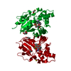 6g22C  6g2lC 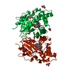 6g2mC  4yihS C: citing same article ( S: Starting model for refinement |
|---|---|
| Similar structure data |


-Links
 PDBj
PDBj- Assembly
| Deposited unit | 
| ||||||||
|---|---|---|---|---|---|---|---|---|---|
| 1 |
| ||||||||
| Unit cell |
|
-Components
| #1: Protein | Mass: 23870.262 Da / Num. of mol.: 2 Source method: isolated from a genetically manipulated source Details: CME is beta-mercapto ethanol covalently bound to cystein residue Source: (gene. exp.) Homo sapiens (human) / Gene: NT5C, DNT1, UMPH2 / Production host:  References: UniProt: Q8TCD5, Hydrolases; Acting on ester bonds; Phosphoric-monoester hydrolases #2: Chemical |   Mass: 24.305 Da / Num. of mol.: 2 / Source method: obtained synthetically / Formula: Mg Mass: 24.305 Da / Num. of mol.: 2 / Source method: obtained synthetically / Formula: Mg#3: Chemical | 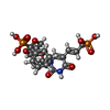  Mass: 516.332 Da / Num. of mol.: 2 / Source method: obtained synthetically / Formula: C19H22N2O11P2 Mass: 516.332 Da / Num. of mol.: 2 / Source method: obtained synthetically / Formula: C19H22N2O11P2#4: Water | ChemComp-HOH / |  Mass: 18.015 Da / Num. of mol.: 291 / Source method: isolated from a natural source / Formula: H2O Mass: 18.015 Da / Num. of mol.: 291 / Source method: isolated from a natural source / Formula: H2OHas protein modification | Y | |
|---|
-Experimental details
-Experiment
| Experiment | Method: X-RAY DIFFRACTION / Number of used crystals: 1 |
|---|
- Sample preparation
| Crystal | Density Matthews: 2.16 Å3/Da / Density % sol: 42.99 % |
|---|---|
| Crystal grow | Temperature: 291 K / Method: vapor diffusion / pH: 8 Details: Morpheus H2, 0.1M MES/imidazole pH 6.5, 10% PEG 8000, 20% ethylene glycol, 0.02M of each amino acid, |
-Data collection
| Diffraction | Mean temperature: 100 K |
|---|---|
| Diffraction source | Source: SYNCHROTRON / Site: BESSY  / Beamline: 14.1 / Wavelength: 0.9184 Å / Beamline: 14.1 / Wavelength: 0.9184 Å |
| Detector | Type: DECTRIS PILATUS 6M / Detector: PIXEL / Date: Nov 15, 2016 |
| Radiation | Protocol: SINGLE WAVELENGTH / Monochromatic (M) / Laue (L): M / Scattering type: x-ray |
| Radiation wavelength | Wavelength: 0.9184 Å / Relative weight: 1 |
| Reflection | Resolution: 1.4→42.41 Å / Num. obs: 68116 / % possible obs: 87.8 % / Redundancy: 2.22 % / Biso Wilson estimate: 27.42 Å2 / CC1/2: 1 / Rrim(I) all: 0.026 / Net I/σ(I): 18.84 |
| Reflection shell | Resolution: 1.4→1.49 Å / Redundancy: 2.21 % / Num. unique obs: 11048 / CC1/2: 0.7414 / Rrim(I) all: 0.556 / % possible all: 87.9 |
-Phasing
| Phasing | Method: molecular replacement | ||||||
|---|---|---|---|---|---|---|---|
| Phasing MR | R rigid body: 0.546
|
- Processing
| Software |
| |||||||||||||||||||||||||||||||||||||||||||||||||||||||||||||||||||||||||||
|---|---|---|---|---|---|---|---|---|---|---|---|---|---|---|---|---|---|---|---|---|---|---|---|---|---|---|---|---|---|---|---|---|---|---|---|---|---|---|---|---|---|---|---|---|---|---|---|---|---|---|---|---|---|---|---|---|---|---|---|---|---|---|---|---|---|---|---|---|---|---|---|---|---|---|---|---|
| Refinement | Method to determine structure: MOLECULAR REPLACEMENT Starting model: 4YIH Resolution: 1.4→42.41 Å / Cor.coef. Fo:Fc: 0.981 / Cor.coef. Fo:Fc free: 0.973 / SU B: 3.966 / SU ML: 0.063 / SU R Cruickshank DPI: 0.0712 / Cross valid method: THROUGHOUT / σ(F): 0 / ESU R: 0.071 / ESU R Free: 0.066 Details: HYDROGENS HAVE BEEN ADDED IN THE RIDING POSITIONS U VALUES : REFINED INDIVIDUALLY
| |||||||||||||||||||||||||||||||||||||||||||||||||||||||||||||||||||||||||||
| Solvent computation | Ion probe radii: 0.8 Å / Shrinkage radii: 0.8 Å / VDW probe radii: 1.2 Å | |||||||||||||||||||||||||||||||||||||||||||||||||||||||||||||||||||||||||||
| Displacement parameters | Biso max: 97.88 Å2 / Biso mean: 27.295 Å2 / Biso min: 14.5 Å2
| |||||||||||||||||||||||||||||||||||||||||||||||||||||||||||||||||||||||||||
| Refinement step | Cycle: final / Resolution: 1.4→42.41 Å
| |||||||||||||||||||||||||||||||||||||||||||||||||||||||||||||||||||||||||||
| Refine LS restraints |
| |||||||||||||||||||||||||||||||||||||||||||||||||||||||||||||||||||||||||||
| LS refinement shell | Resolution: 1.404→1.441 Å / Rfactor Rfree error: 0 / Total num. of bins used: 20
|
