 Movie
Movie Controller

[English] 日本語
 Yorodumi
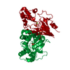
- PDB-4e88: CRYSTAL STRUCTURE OF DE NOVO DESIGNED CYSTEINE ESTERASE ECH13, No... -
+ Open data

- Basic information
| Entry | Database: PDB / ID: 4.0E+88 | ||||||
|---|---|---|---|---|---|---|---|
| Title | CRYSTAL STRUCTURE OF DE NOVO DESIGNED CYSTEINE ESTERASE ECH13, Northeast Structural Genomics Consortium Target OR51 | ||||||
 Components Components | DE NOVO DESIGNED CYSTEINE ESTERASE ECH13 | ||||||
 Keywords Keywords | HYDROLASE / Structural Genomics / PSI-Biology / Protein Structure Initiative / Northeast Structural Genomics Consortium / NESG | ||||||
| Function / homology | Deoxyribonucleotidase; domain 2 / Ribonucleotide Reductase Protein R1; domain 1 / HAD superfamily/HAD-like / Rossmann fold / Orthogonal Bundle / 3-Layer(aba) Sandwich / Mainly Alpha / Alpha Beta Function and homology information Function and homology information | ||||||
| Biological species | synthetic construct (others) | ||||||
| Method |  X-RAY DIFFRACTION / MIR / Resolution: 2 Å X-RAY DIFFRACTION / MIR / Resolution: 2 Å | ||||||
 Authors Authors | Kuzin, A. / Su, M. / Seetharaman, J. / Xiao, X. / Sahdev, S. / Ciccosanti, C. / Richter, F. / Everett, J.K. / Acton, T.B. / Montelione, G.T. ...Kuzin, A. / Su, M. / Seetharaman, J. / Xiao, X. / Sahdev, S. / Ciccosanti, C. / Richter, F. / Everett, J.K. / Acton, T.B. / Montelione, G.T. / Hunt, J.F. / Tong, L. / Northeast Structural Genomics Consortium (NESG) | ||||||
 Citation Citation | Journal: To be Published Title: Northeast Structural Genomics Consortium Target OR51 Authors: Kuzin, A. / Su, M. / Seetharaman, J. / Xiao, X. / Sahdev, S. / Ciccosanti, C. / Richter, F. / Everett, J.K. / Acton, T.B. / Montelione, G.T. / Hunt, J.F. / Tong, L. | ||||||
| History |
|
- Structure visualization
| Structure viewer | Molecule: MolmilJmol/JSmol |
|---|
- Downloads & links
-Download
| PDBx/mmCIF format | 4e88.cif.gz | 103.5 KB | Display | PDBx/mmCIF format |
|---|---|---|---|---|
| PDB format | pdb4e88.ent.gz | 79.2 KB | Display | PDB format |
| PDBx/mmJSON format | 4e88.json.gz | Tree view | PDBx/mmJSON format | |
| Others |  Other downloads Other downloads |
-Validation report
| Arichive directory | https://data.pdbj.org/pub/pdb/validation_reports/e8/4e88ftp://data.pdbj.org/pub/pdb/validation_reports/e8/4e88 | HTTPS FTP |
|---|
-Related structure data
| Related structure data |  3u13S S: Starting model for refinement |
|---|---|
| Similar structure data | |
| Other databases |


-Links
 PDBj
PDBj- Assembly
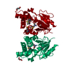
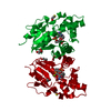
| Deposited unit | 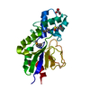
| ||||||||
|---|---|---|---|---|---|---|---|---|---|
| 1 | 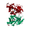
| ||||||||
| Unit cell |
| ||||||||
| Components on special symmetry positions |
| ||||||||
| Details | dimer,54.06 kD,98.7% |
-Components
| #1: Protein | Mass: 24209.996 Da / Num. of mol.: 1 Source method: isolated from a genetically manipulated source Source: (gene. exp.) synthetic construct (others) |
|---|---|
| #2: Chemical | ChemComp-GOL /   Mass: 92.094 Da / Num. of mol.: 1 / Source method: obtained synthetically / Formula: C3H8O3 Mass: 92.094 Da / Num. of mol.: 1 / Source method: obtained synthetically / Formula: C3H8O3 |
| #3: Water | ChemComp-HOH /  Mass: 18.015 Da / Num. of mol.: 183 / Source method: isolated from a natural source / Formula: H2O Mass: 18.015 Da / Num. of mol.: 183 / Source method: isolated from a natural source / Formula: H2O |
| Has protein modification | Y |
-Experimental details
-Experiment
| Experiment | Method: X-RAY DIFFRACTION / Number of used crystals: 1 |
|---|
- Sample preparation
| Crystal | Density Matthews: 2.59 Å3/Da / Density % sol: 52.53 % |
|---|---|
| Crystal grow | Temperature: 277 K / Method: vapor diffusion, sitting drop / pH: 7.5 Details: Protein solution: 100mM NaCl, 5mM DTT, 0.02% NaN3, 10mM Tris-HCl (pH 7.5) . Reservoir solution:100MM NACL, 5MM DTT, 0.02% NAN3, 10MM TRIS-HCL (PH 7.5) . RESERVOIR SOLUTION: 0.2M REMARK 280 ...Details: Protein solution: 100mM NaCl, 5mM DTT, 0.02% NaN3, 10mM Tris-HCl (pH 7.5) . Reservoir solution:100MM NACL, 5MM DTT, 0.02% NAN3, 10MM TRIS-HCL (PH 7.5) . RESERVOIR SOLUTION: 0.2M REMARK 280 NH4F, 20% PEG3350, REMARK 280 0.02% NAN3, 10MM TRIS-HCL (PH 7.5) . RESERVOIR SOLUTION: 0.2M , VAPOR DIFFUSION, SITTING DROP, temperature 277KK |
-Data collection
| Diffraction | Mean temperature: 100 K |
|---|---|
| Diffraction source | Source: ROTATING ANODE / Type: RIGAKU / Wavelength: 1.5418 Å |
| Detector | Type: ADSC QUANTUM 4 / Detector: CCD / Date: May 19, 2011 |
| Radiation | Monochromator: Si 111 CHANNEL / Protocol: SINGLE WAVELENGTH / Monochromatic (M) / Laue (L): M / Scattering type: x-ray |
| Radiation wavelength | Wavelength: 1.5418 Å / Relative weight: 1 |
| Reflection | Resolution: 2→30 Å / Num. obs: 16356 / % possible obs: 97.3 % / Observed criterion σ(I): -3 / Redundancy: 3.8 % / Biso Wilson estimate: 28.24 Å2 / Rmerge(I) obs: 0.046 / Net I/σ(I): 30.9 |
- Processing
| Software |
| |||||||||||||||||||||||||||||||||||||||||||||||||
|---|---|---|---|---|---|---|---|---|---|---|---|---|---|---|---|---|---|---|---|---|---|---|---|---|---|---|---|---|---|---|---|---|---|---|---|---|---|---|---|---|---|---|---|---|---|---|---|---|---|---|
| Refinement | Method to determine structure: MIR Starting model: pdb entry 3U13 Resolution: 2→23.091 Å / Occupancy max: 1 / Occupancy min: 0.38 / FOM work R set: 0.866 / SU ML: 0.28 / Cross valid method: THROUGHOUT / σ(F): 1.38 / Phase error: 20.55 / Stereochemistry target values: ML
| |||||||||||||||||||||||||||||||||||||||||||||||||
| Solvent computation | Shrinkage radii: 0.98 Å / VDW probe radii: 1.2 Å / Solvent model: FLAT BULK SOLVENT MODEL / Bsol: 39.488 Å2 / ksol: 0.322 e/Å3 | |||||||||||||||||||||||||||||||||||||||||||||||||
| Displacement parameters | Biso max: 84.78 Å2 / Biso mean: 33.286 Å2 / Biso min: 13.95 Å2
| |||||||||||||||||||||||||||||||||||||||||||||||||
| Refinement step | Cycle: LAST / Resolution: 2→23.091 Å
| |||||||||||||||||||||||||||||||||||||||||||||||||
| Refine LS restraints |
| |||||||||||||||||||||||||||||||||||||||||||||||||
| LS refinement shell | Refine-ID: X-RAY DIFFRACTION / Total num. of bins used: 6
| |||||||||||||||||||||||||||||||||||||||||||||||||
| Refinement TLS params. | Method: refined / Origin x: 10.9111 Å / Origin y: -31.222 Å / Origin z: -10.3695 Å
| |||||||||||||||||||||||||||||||||||||||||||||||||
| Refinement TLS group |
|
