| Entry | Database: PDB / ID: 2jao
|
|---|
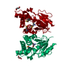
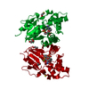
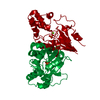
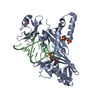
| Title | Crystal structure of D12N variant of mouse cytosolic 5'(3')- deoxyribonucleotidase (cdN) in complex with deoxyguanosine 5'- monophosphate |
|---|
 Components Components | 5'(3')-DEOXYRIBONUCLEOTIDASE |
|---|
 Keywords Keywords | HYDROLASE / NUCLEOTIDE METABOLISM / NUCLEOTIDE-BINDING / ALPHA-BETA FOLD / METAL-BINDING / MAGNESIUM / CYTOSOL |
|---|
| Function / homology |  Function and homology information Function and homology information
Purine catabolism / pyrimidine nucleotide binding / Pyrimidine catabolism / pyrimidine deoxyribonucleotide catabolic process / dTMP catabolic process / dCMP catabolic process / nucleotidase activity / dUMP catabolic process / dGMP catabolic process / IMP catabolic process ...Purine catabolism / pyrimidine nucleotide binding / Pyrimidine catabolism / pyrimidine deoxyribonucleotide catabolic process / dTMP catabolic process / dCMP catabolic process / nucleotidase activity / dUMP catabolic process / dGMP catabolic process / IMP catabolic process / deoxyribonucleotide catabolic process / Hydrolases; Acting on ester bonds; Phosphoric-monoester hydrolases / 5'-nucleotidase activity / phosphatase activity / mitochondrion / metal ion binding / identical protein binding / nucleus / cytoplasm / cytosolSimilarity search - Function 5'(3')-deoxyribonucleotidase / 5' nucleotidase, deoxy (Pyrimidine), cytosolic type C protein (NT5C) / Deoxyribonucleotidase; domain 2 / Ribonucleotide Reductase Protein R1; domain 1 / HAD superfamily/HAD-like / HAD superfamily / HAD-like superfamily / Rossmann fold / Orthogonal Bundle / 3-Layer(aba) Sandwich ...5'(3')-deoxyribonucleotidase / 5' nucleotidase, deoxy (Pyrimidine), cytosolic type C protein (NT5C) / Deoxyribonucleotidase; domain 2 / Ribonucleotide Reductase Protein R1; domain 1 / HAD superfamily/HAD-like / HAD superfamily / HAD-like superfamily / Rossmann fold / Orthogonal Bundle / 3-Layer(aba) Sandwich / Mainly Alpha / Alpha BetaSimilarity search - Domain/homology |
|---|
| Biological species |   MUS MUSCULUS (house mouse) MUS MUSCULUS (house mouse) |
|---|
| Method |  X-RAY DIFFRACTION / SYNCHROTRON / MOLECULAR REPLACEMENT / Resolution: 2 Å X-RAY DIFFRACTION / SYNCHROTRON / MOLECULAR REPLACEMENT / Resolution: 2 Å |
|---|
 Authors Authors | Wallden, K. / Ruzzenente, B. / Bianchi, V. / Nordlund, P. |
|---|
 Citation Citation | Journal: Biochemistry / Year: 2007
Title: Crystal Structures of Human and Murine Deoxyribonucleotidases: Insights Into Recognition of Substrates and Nucleotide Analogues.
Authors: Wallden, K. / Rinaldo-Matthis, A. / Ruzzenente, B. / Rampazzo, C. / Bianchi, V. / Nordlund, P. |
|---|
| History | | Deposition | Nov 29, 2006 | Deposition site: PDBE / Processing site: PDBE |
|---|
| Revision 1.0 | Nov 20, 2007 | Provider: repository / Type: Initial release |
|---|
| Revision 1.1 | May 8, 2011 | Group: Version format compliance |
|---|
| Revision 1.2 | Jul 13, 2011 | Group: Version format compliance |
|---|
| Revision 1.3 | Jan 17, 2018 | Group: Data collection / Category: diffrn_source / Item: _diffrn_source.pdbx_synchrotron_site |
|---|
| Revision 1.4 | May 15, 2019 | Group: Data collection / Derived calculations / Experimental preparation
Category: chem_comp / exptl_crystal_grow ...chem_comp / exptl_crystal_grow / pdbx_struct_special_symmetry / struct_biol
Item: _chem_comp.type / _exptl_crystal_grow.method / _exptl_crystal_grow.temp |
|---|
| Revision 1.5 | May 8, 2024 | Group: Data collection / Database references ...Data collection / Database references / Derived calculations / Other
Category: chem_comp_atom / chem_comp_bond ...chem_comp_atom / chem_comp_bond / database_2 / pdbx_database_status / pdbx_struct_conn_angle / struct_conn / struct_site
Item: _database_2.pdbx_DOI / _database_2.pdbx_database_accession ..._database_2.pdbx_DOI / _database_2.pdbx_database_accession / _pdbx_database_status.status_code_sf / _pdbx_struct_conn_angle.ptnr1_auth_comp_id / _pdbx_struct_conn_angle.ptnr1_auth_seq_id / _pdbx_struct_conn_angle.ptnr1_label_alt_id / _pdbx_struct_conn_angle.ptnr1_label_asym_id / _pdbx_struct_conn_angle.ptnr1_label_atom_id / _pdbx_struct_conn_angle.ptnr1_label_comp_id / _pdbx_struct_conn_angle.ptnr1_label_seq_id / _pdbx_struct_conn_angle.ptnr3_auth_comp_id / _pdbx_struct_conn_angle.ptnr3_auth_seq_id / _pdbx_struct_conn_angle.ptnr3_label_alt_id / _pdbx_struct_conn_angle.ptnr3_label_asym_id / _pdbx_struct_conn_angle.ptnr3_label_atom_id / _pdbx_struct_conn_angle.ptnr3_label_comp_id / _pdbx_struct_conn_angle.ptnr3_label_seq_id / _pdbx_struct_conn_angle.value / _struct_conn.pdbx_dist_value / _struct_conn.pdbx_ptnr1_label_alt_id / _struct_conn.ptnr1_auth_comp_id / _struct_conn.ptnr1_auth_seq_id / _struct_conn.ptnr1_label_asym_id / _struct_conn.ptnr1_label_atom_id / _struct_conn.ptnr1_label_comp_id / _struct_conn.ptnr1_label_seq_id / _struct_conn.ptnr2_auth_comp_id / _struct_conn.ptnr2_auth_seq_id / _struct_conn.ptnr2_label_asym_id / _struct_conn.ptnr2_label_atom_id / _struct_conn.ptnr2_label_comp_id / _struct_conn.ptnr2_label_seq_id / _struct_site.pdbx_auth_asym_id / _struct_site.pdbx_auth_comp_id / _struct_site.pdbx_auth_seq_id |
|---|
|
|---|
 Movie
Movie Controller

 Yorodumi
 Open data
 Basic information
 Structure visualization
 Downloads & links
 Other downloads
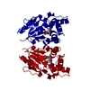


 PDBj
PDBj Assembly


 Mass: 92.094 Da / Num. of mol.: 1 / Source method: obtained synthetically / Formula: C3H8O3
 Mass: 347.221 Da / Num. of mol.: 1 / Source method: obtained synthetically / Formula: C10H14N5O7P
 Mass: 24.305 Da / Num. of mol.: 1 / Source method: obtained synthetically / Formula: Mg
 Mass: 18.015 Da / Num. of mol.: 175 / Source method: isolated from a natural source / Formula: H2O
 Sample preparation
 / Beamline: I711 / Wavelength: 1.141
  Processing