 Movie
Movie Controller

[English] 日本語
 Yorodumi
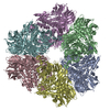
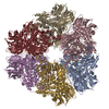
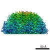
- PDB-6qxm: Cryo-EM structure of T7 bacteriophage portal protein, 12mer, open... -
+ Open data

- Basic information
| Entry | Database: PDB / ID: 6qxm | ||||||||||||||||||||||||
|---|---|---|---|---|---|---|---|---|---|---|---|---|---|---|---|---|---|---|---|---|---|---|---|---|---|
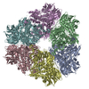
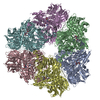
| Title | Cryo-EM structure of T7 bacteriophage portal protein, 12mer, open valve | ||||||||||||||||||||||||
 Components Components | Portal protein | ||||||||||||||||||||||||
 Keywords Keywords | VIRAL PROTEIN / DNA packaging | ||||||||||||||||||||||||
| Function / homology | Portal protein, Caudovirales / Head-to-tail connector protein, podovirus-type / Bacteriophage head to tail connecting protein / viral portal complex / symbiont genome ejection through host cell envelope, short tail mechanism / viral DNA genome packaging / Portal protein Function and homology information Function and homology information | ||||||||||||||||||||||||
| Biological species |   Enterobacteria phage T7 (virus) Enterobacteria phage T7 (virus) | ||||||||||||||||||||||||
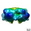
| Method | ELECTRON MICROSCOPY / single particle reconstruction / cryo EM / Resolution: 4.1 Å | ||||||||||||||||||||||||
 Authors Authors | Fabrega-Ferrer, M. / Cuervo, A. / Machon, C. / Fernandez, F.J. / Perez-Luque, R. / Pous, J. / Vega, M.C. / Carrascosa, J.L. / Coll, M. | ||||||||||||||||||||||||
| Funding support |  Spain, 7items Spain, 7items
| ||||||||||||||||||||||||
 Citation Citation | Journal: Nat Commun / Year: 2019 Title: Structures of T7 bacteriophage portal and tail suggest a viral DNA retention and ejection mechanism. Authors: Ana Cuervo / Montserrat Fàbrega-Ferrer / Cristina Machón / José Javier Conesa / Francisco J Fernández / Rosa Pérez-Luque / Mar Pérez-Ruiz / Joan Pous / M Cristina Vega / José L Carrascosa / Miquel Coll / Abstract: Double-stranded DNA bacteriophages package their genome at high pressure inside a procapsid through the portal, an oligomeric ring protein located at a unique capsid vertex. Once the DNA has been ...Double-stranded DNA bacteriophages package their genome at high pressure inside a procapsid through the portal, an oligomeric ring protein located at a unique capsid vertex. Once the DNA has been packaged, the tail components assemble on the portal to render the mature infective virion. The tail tightly seals the ejection conduit until infection, when its interaction with the host membrane triggers the opening of the channel and the viral genome is delivered to the host cell. Using high-resolution cryo-electron microscopy and X-ray crystallography, here we describe various structures of the T7 bacteriophage portal and fiber-less tail complex, which suggest a possible mechanism for DNA retention and ejection: a portal closed conformation temporarily retains the genome before the tail is assembled, whereas an open portal is found in the tail. Moreover, a fold including a seven-bladed β-propeller domain is described for the nozzle tail protein. | ||||||||||||||||||||||||
| History |
|
- Structure visualization
| Movie |
Movie viewer |
|---|---|
| Structure viewer | Molecule: MolmilJmol/JSmol |
- Downloads & links
-Download
| PDBx/mmCIF format | 6qxm.cif.gz | 806.2 KB | Display | PDBx/mmCIF format |
|---|---|---|---|---|
| PDB format | pdb6qxm.ent.gz | 672.5 KB | Display | PDB format |
| PDBx/mmJSON format | 6qxm.json.gz | Tree view | PDBx/mmJSON format | |
| Others |  Other downloads Other downloads |
-Validation report
| Arichive directory | https://data.pdbj.org/pub/pdb/validation_reports/qx/6qxmftp://data.pdbj.org/pub/pdb/validation_reports/qx/6qxm | HTTPS FTP |
|---|
-Related structure data
| Related structure data | 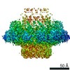 4669MC 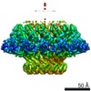 4667C  4706C 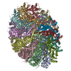 6qwpC 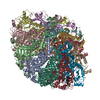 6qx5C 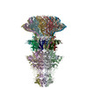 6r21C C: citing same article ( M: map data used to model this data |
|---|---|
| Similar structure data |

-Links
 PDBj
PDBj- Assembly
| Deposited unit | 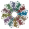
|
|---|---|
| 1 |
|
-Components
| #1: Protein | Mass: 60458.387 Da / Num. of mol.: 12 Source method: isolated from a genetically manipulated source Source: (gene. exp.) Enterobacteria phage T7 (virus) / Production host:  |
|---|
-Experimental details
-Experiment
| Experiment | Method: ELECTRON MICROSCOPY |
|---|---|
| EM experiment | Aggregation state: PARTICLE / 3D reconstruction method: single particle reconstruction |
- Sample preparation
| Component | Name: Bacteriophage packaging protein / Type: COMPLEX / Entity ID: all / Source: RECOMBINANT |
|---|---|
| Source (natural) | Organism: Enterobacteria phage T7 (virus) |
| Source (recombinant) | Organism: |
| Buffer solution | pH: 7.8 |
| Specimen | Embedding applied: NO / Shadowing applied: NO / Staining applied: NO / Vitrification applied: YES |
| Vitrification | Cryogen name: ETHANE |
- Electron microscopy imaging
| Experimental equipment |  Model: Titan Krios / Image courtesy: FEI Company |
|---|---|
| Microscopy | Model: FEI TITAN KRIOS |
| Electron gun | Electron source:  FIELD EMISSION GUN / Accelerating voltage: 300 kV / Illumination mode: FLOOD BEAM FIELD EMISSION GUN / Accelerating voltage: 300 kV / Illumination mode: FLOOD BEAM |
| Electron lens | Mode: BRIGHT FIELD |
| Image recording | Electron dose: 39.4 e/Å2 / Film or detector model: GATAN K2 QUANTUM (4k x 4k) |
- Processing
| CTF correction | Type: PHASE FLIPPING AND AMPLITUDE CORRECTION |
|---|---|
| Symmetry | Point symmetry: C12 (12 fold cyclic) |
| 3D reconstruction | Resolution: 4.1 Å / Resolution method: FSC 0.143 CUT-OFF / Num. of particles: 32688 / Symmetry type: POINT |
