 Movie
Movie Controller


+ Open data

- Basic information
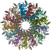
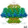
| Entry | Database: EMDB / ID: EMD-4669 | ||||||||||||||||||||||||
|---|---|---|---|---|---|---|---|---|---|---|---|---|---|---|---|---|---|---|---|---|---|---|---|---|---|
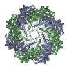
| Title | Bacteriophage packaging protein | ||||||||||||||||||||||||
 Map data Map data | |||||||||||||||||||||||||
 Sample Sample |
| ||||||||||||||||||||||||
 Keywords Keywords | viral protein / DNA packaging | ||||||||||||||||||||||||
| Function / homology | Portal protein, Caudovirales / Head-to-tail connector protein, podovirus-type / Bacteriophage head to tail connecting protein / viral portal complex / symbiont genome ejection through host cell envelope, short tail mechanism / viral DNA genome packaging / Portal protein Function and homology information Function and homology information | ||||||||||||||||||||||||
| Biological species |   Enterobacteria phage T7 (virus) Enterobacteria phage T7 (virus) | ||||||||||||||||||||||||
| Method | single particle reconstruction / cryo EM / Resolution: 4.1 Å | ||||||||||||||||||||||||
 Authors Authors | Fabrega-Ferrer M / Cuervo A | ||||||||||||||||||||||||
| Funding support |  Spain, 7 items Spain, 7 items
| ||||||||||||||||||||||||
 Citation Citation | Journal: Nat Commun / Year: 2019 Title: Structures of T7 bacteriophage portal and tail suggest a viral DNA retention and ejection mechanism. Authors: Ana Cuervo / Montserrat Fàbrega-Ferrer / Cristina Machón / José Javier Conesa / Francisco J Fernández / Rosa Pérez-Luque / Mar Pérez-Ruiz / Joan Pous / M Cristina Vega / José L Carrascosa / Miquel Coll / Abstract: Double-stranded DNA bacteriophages package their genome at high pressure inside a procapsid through the portal, an oligomeric ring protein located at a unique capsid vertex. Once the DNA has been ...Double-stranded DNA bacteriophages package their genome at high pressure inside a procapsid through the portal, an oligomeric ring protein located at a unique capsid vertex. Once the DNA has been packaged, the tail components assemble on the portal to render the mature infective virion. The tail tightly seals the ejection conduit until infection, when its interaction with the host membrane triggers the opening of the channel and the viral genome is delivered to the host cell. Using high-resolution cryo-electron microscopy and X-ray crystallography, here we describe various structures of the T7 bacteriophage portal and fiber-less tail complex, which suggest a possible mechanism for DNA retention and ejection: a portal closed conformation temporarily retains the genome before the tail is assembled, whereas an open portal is found in the tail. Moreover, a fold including a seven-bladed β-propeller domain is described for the nozzle tail protein. | ||||||||||||||||||||||||
| History |
|
- Structure visualization
| Movie |
Movie viewer |
|---|---|
| Structure viewer | EM map: SurfViewMolmilJmol/JSmol |
| Supplemental images |

- Downloads & links
-EMDB archive
| Map data | emd_4669.map.gz | 15.2 MB | EMDB map data format | |
|---|---|---|---|---|
| Header (meta data) | emd-4669-v30.xmlemd-4669.xml | 11.5 KB 11.5 KB | Display Display | EMDB header |
| FSC (resolution estimation) | emd_4669_fsc.xml | 5.5 KB | Display | FSC data file |
| Images | 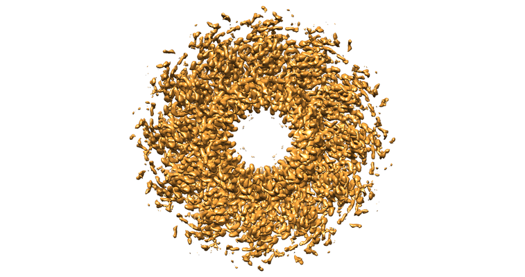 emd_4669.png emd_4669.png | 248.9 KB | ||
| Filedesc metadata | emd-4669.cif.gz | 5.7 KB | ||
| Archive directory |  http://ftp.pdbj.org/pub/emdb/structures/EMD-4669ftp://ftp.pdbj.org/pub/emdb/structures/EMD-4669 http://ftp.pdbj.org/pub/emdb/structures/EMD-4669ftp://ftp.pdbj.org/pub/emdb/structures/EMD-4669 | HTTPS FTP |
-Related structure data
| Related structure data |  6qxmMC  4667C  4706C  6qwpC 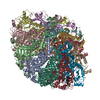 6qx5C  6r21C C: citing same article ( M: atomic model generated by this map |
|---|---|
| Similar structure data |


-Links
| EMDB pages | EMDB (EBI/PDBe) / EMDataResource |
|---|
-Map
| File | Download / File: emd_4669.map.gz / Format: CCP4 / Size: 40.6 MB / Type: IMAGE STORED AS FLOATING POINT NUMBER (4 BYTES) | ||||||||||||||||||||||||||||||||||||||||||||||||||||||||||||||||||||
|---|---|---|---|---|---|---|---|---|---|---|---|---|---|---|---|---|---|---|---|---|---|---|---|---|---|---|---|---|---|---|---|---|---|---|---|---|---|---|---|---|---|---|---|---|---|---|---|---|---|---|---|---|---|---|---|---|---|---|---|---|---|---|---|---|---|---|---|---|---|
| Projections & slices | Image control
Images are generated by Spider. | ||||||||||||||||||||||||||||||||||||||||||||||||||||||||||||||||||||
| Voxel size | X=Y=Z: 1.04 Å | ||||||||||||||||||||||||||||||||||||||||||||||||||||||||||||||||||||
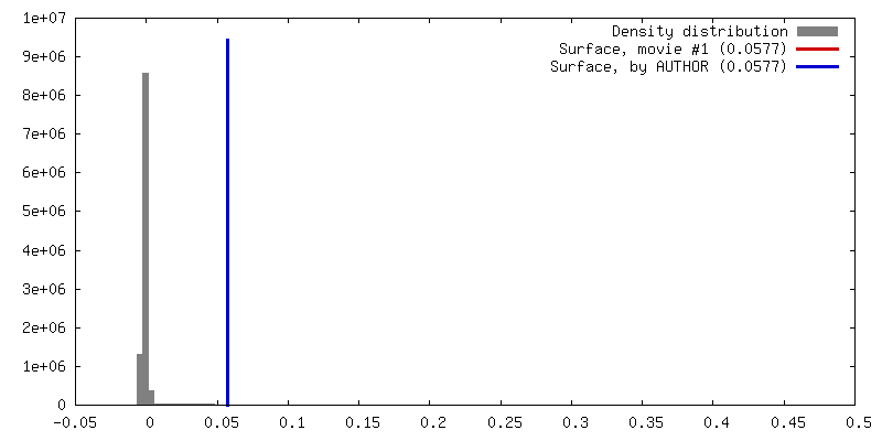
| Density |
| ||||||||||||||||||||||||||||||||||||||||||||||||||||||||||||||||||||
| Symmetry | Space group: 1 | ||||||||||||||||||||||||||||||||||||||||||||||||||||||||||||||||||||
| Details | EMDB XML:
CCP4 map header:
| ||||||||||||||||||||||||||||||||||||||||||||||||||||||||||||||||||||
 Z (Sec.)
 Y (Row.)
 X (Col.)


-Supplemental data
- Sample components
-Entire : Bacteriophage packaging protein
| Entire | Name: Bacteriophage packaging protein |
|---|---|
| Components |
|
-Supramolecule #1: Bacteriophage packaging protein
| Supramolecule | Name: Bacteriophage packaging protein / type: complex / ID: 1 / Parent: 0 / Macromolecule list: all |
|---|---|
| Source (natural) | Organism: Enterobacteria phage T7 (virus) |
-Macromolecule #1: Portal protein
| Macromolecule | Name: Portal protein / type: protein_or_peptide / ID: 1 / Number of copies: 12 / Enantiomer: LEVO |
|---|---|
| Source (natural) | Organism: Enterobacteria phage T7 (virus) |
| Molecular weight | Theoretical: 60.458387 KDa |
| Recombinant expression | Organism:  |
| Sequence | String: MAEKRTGLAE DGAKSVYERL KNDRAPYETR AQNCAQYTIP SLFPKDSDNA STDYQTPWQA VGARGLNNLA SKLMLALFPM QTWMRLTIS EYEAKQLLSD PDGLAKVDEG LSMVERIIMN YIESNSYRVT LFEALKQLVV AGNVLLYLPE PEGSNYNPMK L YRLSSYVV ...String: MAEKRTGLAE DGAKSVYERL KNDRAPYETR AQNCAQYTIP SLFPKDSDNA STDYQTPWQA VGARGLNNLA SKLMLALFPM QTWMRLTIS EYEAKQLLSD PDGLAKVDEG LSMVERIIMN YIESNSYRVT LFEALKQLVV AGNVLLYLPE PEGSNYNPMK L YRLSSYVV QRDAFGNVLQ MVTRDQIAFG ALPEDIRKAV EGQGGEKKAD ETIDVYTHIY LDEDSGEYLR YEEVEGMEVQ GS DGTYPKE ACPYIPIRMV RLDGESYGRS YIEEYLGDLR SLENLQEAIV KMSMISSKVI GLVNPAGITQ PRRLTKAQTG DFV TGRPED ISFLQLEKQA DFTVAKAVSD AIEARLSFAF MLNSAVQRTG ERVTAEEIRY VASELEDTLG GVYSILSQEL QLPL VRVLL KQLQATQQIP ELPKEAVEPT ISTGLEAIGR GQDLDKLERC VTAWAALAPM RDDPDINLAM IKLRIANAIG IDTSG ILLT EEQKQQKMAQ QSMQMGMDNG AAALAQGMAA QATASPEAMA AAADSVGLQP GIAAALEHHH HHH UniProtKB: Portal protein |
-Experimental details
-Structure determination
| Method | cryo EM |
|---|---|
 Processing Processing | single particle reconstruction |
| Aggregation state | particle |
-Sample preparation
| Buffer | pH: 7.8 |
|---|---|
| Vitrification | Cryogen name: ETHANE |
- Electron microscopy
| Microscope | FEI TITAN KRIOS |
|---|---|
| Image recording | Film or detector model: GATAN K2 QUANTUM (4k x 4k) / Average electron dose: 39.4 e/Å2 |
| Electron beam | Acceleration voltage: 300 kV / Electron source:  FIELD EMISSION GUN FIELD EMISSION GUN |
| Electron optics | Illumination mode: FLOOD BEAM / Imaging mode: BRIGHT FIELD |
| Experimental equipment |  Model: Titan Krios / Image courtesy: FEI Company |

