Movie
Movie Controller

[English] 日本語
 Yorodumi
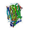
- PDB-6qq5: Cryo-EM structure of dimeric quinol dependent nitric oxide reduct... -
+ Open data

- Basic information
| Entry | Database: PDB / ID: 6qq5 | |||||||||||||||
|---|---|---|---|---|---|---|---|---|---|---|---|---|---|---|---|---|
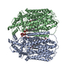
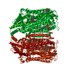
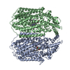
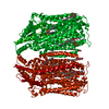
| Title | Cryo-EM structure of dimeric quinol dependent nitric oxide reductase (qNOR) from Alcaligenes xylosoxidans | |||||||||||||||
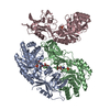
 Components Components | Nitric oxide reductase subunit B | |||||||||||||||
 Keywords Keywords | OXIDOREDUCTASE / Proton Transfer / Membrane Protein / Homodimer | |||||||||||||||
| Function / homology |  Function and homology information Function and homology informationnitric oxide reductase (cytochrome c) / nitric oxide reductase activity / cytochrome-c oxidase activity / aerobic respiration / heme binding / membrane Similarity search - Function | |||||||||||||||
| Biological species |  Alcaligenes xylosoxydans xylosoxydans (bacteria) Alcaligenes xylosoxydans xylosoxydans (bacteria) | |||||||||||||||
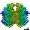
| Method | ELECTRON MICROSCOPY / single particle reconstruction / cryo EM / Resolution: 3.9 Å | |||||||||||||||
 Authors Authors | Gopalasingam, C.C. / Johnson, R.M. / Chiduza, G.N. / Tosha, T. / Yamamoto, M. / Shiro, Y. / Antonyuk, S.V. / Muench, S.P. / Hasnain, S.S. | |||||||||||||||
| Funding support |  United Kingdom, 4items United Kingdom, 4items
| |||||||||||||||
 Citation Citation | Journal: Sci Adv / Year: 2019 Title: Dimeric structures of quinol-dependent nitric oxide reductases (qNORs) revealed by cryo-electron microscopy. Authors: Chai C Gopalasingam / Rachel M Johnson / George N Chiduza / Takehiko Tosha / Masaki Yamamoto / Yoshitsugu Shiro / Svetlana V Antonyuk / Stephen P Muench / S Samar Hasnain /  Abstract: Quinol-dependent nitric oxide reductases (qNORs) are membrane-integrated, iron-containing enzymes of the denitrification pathway, which catalyze the reduction of nitric oxide (NO) to the major ozone ...Quinol-dependent nitric oxide reductases (qNORs) are membrane-integrated, iron-containing enzymes of the denitrification pathway, which catalyze the reduction of nitric oxide (NO) to the major ozone destroying gas nitrous oxide (NO). Cryo-electron microscopy structures of active qNOR from and an activity-enhancing mutant have been determined to be at local resolutions of 3.7 and 3.2 Å, respectively. They unexpectedly reveal a dimeric conformation (also confirmed for qNOR from ) and define the active-site configuration, with a clear water channel from the cytoplasm. Structure-based mutagenesis has identified key residues involved in proton transport and substrate delivery to the active site of qNORs. The proton supply direction differs from cytochrome c-dependent NOR (cNOR), where water molecules from the cytoplasm serve as a proton source similar to those from cytochrome c oxidase. | |||||||||||||||
| History |
|
- Structure visualization
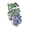
| Movie |
Movie viewer |
|---|---|
| Structure viewer | Molecule: MolmilJmol/JSmol |
- Downloads & links
-Download
| PDBx/mmCIF format | 6qq5.cif.gz | 266 KB | Display | PDBx/mmCIF format |
|---|---|---|---|---|
| PDB format | pdb6qq5.ent.gz | 216.6 KB | Display | PDB format |
| PDBx/mmJSON format | 6qq5.json.gz | Tree view | PDBx/mmJSON format | |
| Others |  Other downloads Other downloads |
-Validation report
| Arichive directory | https://data.pdbj.org/pub/pdb/validation_reports/qq/6qq5ftp://data.pdbj.org/pub/pdb/validation_reports/qq/6qq5 | HTTPS FTP |
|---|
-Related structure data
| Related structure data | 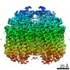 4618MC 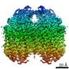 4619C 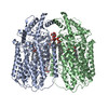 6qq6C M: map data used to model this data C: citing same article ( |
|---|---|
| Similar structure data |


-Links
 PDBj
PDBj

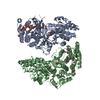
- Assembly
| Deposited unit | 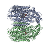
|
|---|---|
| 1 |
|
-Components
| #1: Protein | Mass: 83121.984 Da / Num. of mol.: 2 Source method: isolated from a genetically manipulated source Source: (gene. exp.) Alcaligenes xylosoxydans xylosoxydans (bacteria)Gene: norB_1, ERS451415_02175 / Plasmid: pET-26b (+) / Production host: References: UniProt: A0A0D6H8R3, nitric oxide reductase (cytochrome c) #2: Chemical | ChemComp-HEM /   Mass: 616.487 Da / Num. of mol.: 4 / Source method: obtained synthetically / Formula: C34H32FeN4O4 Mass: 616.487 Da / Num. of mol.: 4 / Source method: obtained synthetically / Formula: C34H32FeN4O4#3: Chemical |   Mass: 55.845 Da / Num. of mol.: 2 / Source method: obtained synthetically / Formula: Fe Mass: 55.845 Da / Num. of mol.: 2 / Source method: obtained synthetically / Formula: Fe#4: Chemical |   Mass: 40.078 Da / Num. of mol.: 2 / Source method: obtained synthetically / Formula: Ca Mass: 40.078 Da / Num. of mol.: 2 / Source method: obtained synthetically / Formula: Ca |
|---|
-Experimental details
-Experiment
| Experiment | Method: ELECTRON MICROSCOPY |
|---|---|
| EM experiment | Aggregation state: PARTICLE / 3D reconstruction method: single particle reconstruction |
- Sample preparation
| Component | Name: Quinol Dependent Nitric Oxide Reductase / Type: COMPLEX / Entity ID: #1 / Source: RECOMBINANT |
|---|---|
| Molecular weight | Value: 0.17 MDa / Experimental value: NO |
| Source (natural) | Organism: Achromobacter xylosoxidans (bacteria) |
| Source (recombinant) | Organism: |
| Buffer solution | pH: 7 |
| Specimen | Conc.: 3 mg/ml / Embedding applied: NO / Shadowing applied: NO / Staining applied: NO / Vitrification applied: YES |
| Vitrification | Instrument: FEI VITROBOT MARK IV / Cryogen name: ETHANE / Humidity: 100 % / Chamber temperature: 277 K Details: Blot for 6 seconds with blot force of 6 prior to plunge freezing |
- Electron microscopy imaging
| Experimental equipment |  Model: Titan Krios / Image courtesy: FEI Company |
|---|---|
| Microscopy | Model: FEI TITAN KRIOS |
| Electron gun | Electron source:  FIELD EMISSION GUN / Accelerating voltage: 300 kV / Illumination mode: FLOOD BEAM FIELD EMISSION GUN / Accelerating voltage: 300 kV / Illumination mode: FLOOD BEAM |
| Electron lens | Mode: BRIGHT FIELD / Nominal magnification: 75000 X / Nominal defocus max: -3500 nm / Nominal defocus min: -1500 nm / Cs: 2.7 mm |
| Specimen holder | Cryogen: NITROGEN / Specimen holder model: FEI TITAN KRIOS AUTOGRID HOLDER |
| Image recording | Electron dose: 65 e/Å2 / Film or detector model: GATAN K2 SUMMIT (4k x 4k) / Num. of real images: 3213 |
| Image scans | Movie frames/image: 40 |
- Processing
| EM software |
| ||||||||||||||||||||||||||||
|---|---|---|---|---|---|---|---|---|---|---|---|---|---|---|---|---|---|---|---|---|---|---|---|---|---|---|---|---|---|
| CTF correction | Type: PHASE FLIPPING AND AMPLITUDE CORRECTION | ||||||||||||||||||||||||||||
| Particle selection | Num. of particles selected: 707246 | ||||||||||||||||||||||||||||
| Symmetry | Point symmetry: C2 (2 fold cyclic) | ||||||||||||||||||||||||||||
| 3D reconstruction | Resolution: 3.9 Å / Resolution method: FSC 0.143 CUT-OFF / Num. of particles: 56134 / Algorithm: FOURIER SPACE / Symmetry type: POINT | ||||||||||||||||||||||||||||
| Atomic model building | Protocol: FLEXIBLE FIT / Space: REAL | ||||||||||||||||||||||||||||
| Atomic model building | PDB-ID: 3AYF Pdb chain-ID: A / Accession code: 3AYF / Source name: PDB / Type: experimental model |