Movie
Movie Controller

[English] 日本語
 Yorodumi
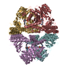
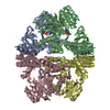
- EMDB-0822: Cryo-EM structure of dimeric quinol dependent Nitric Oxide Reduct... -
+ Open data

- Basic information
| Entry | Database: EMDB / ID: EMD-0822 | |||||||||||||||||||||
|---|---|---|---|---|---|---|---|---|---|---|---|---|---|---|---|---|---|---|---|---|---|---|
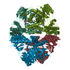
| Title | Cryo-EM structure of dimeric quinol dependent Nitric Oxide Reductase (qNOR) from the pathogen Neisseria meninigitidis | |||||||||||||||||||||
 Map data Map data | ||||||||||||||||||||||
 Sample Sample |
| |||||||||||||||||||||
 Keywords Keywords | Neisseria meningitidis / quinol-dependent electrogenic Nitric Oxide Reductase (qNOR) / OXIDOREDUCTASE | |||||||||||||||||||||
| Function / homology |  Function and homology information Function and homology informationnitric-oxide reductase / cytochrome-c oxidase activity / aerobic respiration / oxidoreductase activity / heme binding / membrane / metal ion binding Similarity search - Function | |||||||||||||||||||||
| Biological species |  Neisseria meningitidis alpha14 (bacteria) Neisseria meningitidis alpha14 (bacteria) | |||||||||||||||||||||
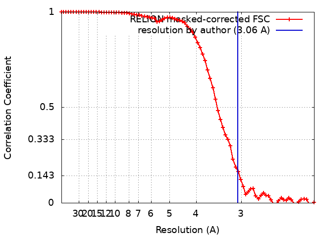
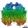
| Method | single particle reconstruction / cryo EM / Resolution: 3.06 Å | |||||||||||||||||||||
 Authors Authors | Jamali MMA / Gopalasingam CC | |||||||||||||||||||||
| Funding support |  Japan, Japan,  United Kingdom, 6 items United Kingdom, 6 items
| |||||||||||||||||||||
 Citation Citation | Journal: IUCrJ / Year: 2020 Title: The active form of quinol-dependent nitric oxide reductase from is a dimer. Authors: M Arif M Jamali / Chai C Gopalasingam / Rachel M Johnson / Takehiko Tosha / Kazumasa Muramoto / Stephen P Muench / Svetlana V Antonyuk / Yoshitsugu Shiro / Samar S Hasnain / Abstract: is carried by nearly a billion humans, causing developmental impairment and over 100 000 deaths a year. A quinol-dependent nitric oxide reductase (qNOR) plays a critical role in the survival of ... is carried by nearly a billion humans, causing developmental impairment and over 100 000 deaths a year. A quinol-dependent nitric oxide reductase (qNOR) plays a critical role in the survival of the bacterium in the human host. X-ray crystallographic analyses of qNOR, including that from (qNOR) reported here at 3.15 Å resolution, show monomeric assemblies, despite the more active dimeric sample being used for crystallization. Cryo-electron microscopic analysis of the same chromatographic fraction of qNOR, however, revealed a dimeric assembly at 3.06 Å resolution. It is shown that zinc (which is used in crystallization) binding near the dimer-stabilizing TMII region contributes to the disruption of the dimer. A similar destabilization is observed in the monomeric (∼85 kDa) cryo-EM structure of a mutant (Glu494Ala) qNOR from the opportunistic pathogen () , which primarily migrates as a monomer. The monomer-dimer transition of qNORs seen in the cryo-EM and crystallographic structures has wider implications for structural studies of multimeric membrane proteins. X-ray crystallographic and cryo-EM structural analyses have been performed on the same chromatographic fraction of qNOR to high resolution. This represents one of the first examples in which the two approaches have been used to reveal a monomeric assembly and a dimeric assembly in vitrified cryo-EM grids. A number of factors have been identified that may trigger the destabilization of helices that are necessary to preserve the integrity of the dimer. These include zinc binding near the entry of the putative proton-transfer channel and the preservation of the conformational integrity of the active site. The mutation near the active site results in disruption of the active site, causing an additional destabilization of helices (TMIX and TMX) that flank the proton-transfer channel helices, creating an inert monomeric enzyme. | |||||||||||||||||||||
| History |
|
- Structure visualization
| Movie |
Movie viewer |
|---|---|
| Structure viewer | EM map: SurfViewMolmilJmol/JSmol |
| Supplemental images |

- Downloads & links
-EMDB archive
| Map data | emd_0822.map.gz | 28.4 MB | EMDB map data format | |
|---|---|---|---|---|
| Header (meta data) | emd-0822-v30.xmlemd-0822.xml | 15.7 KB 15.7 KB | Display Display | EMDB header |
| FSC (resolution estimation) | emd_0822_fsc.xml | 7.2 KB | Display | FSC data file |
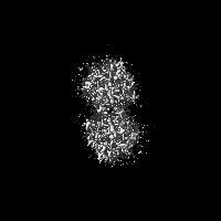
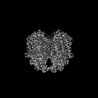
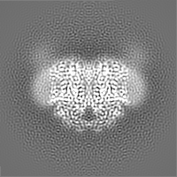
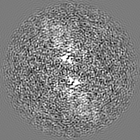
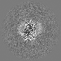
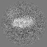
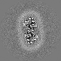
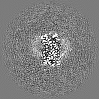
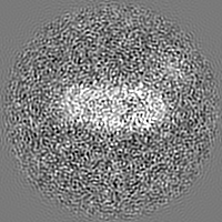
| Images | 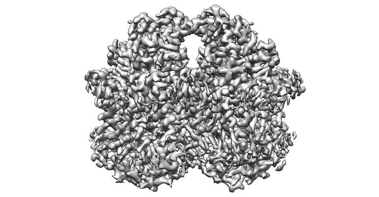 emd_0822.png emd_0822.png | 122.6 KB | ||
| Filedesc metadata | emd-0822.cif.gz | 6.6 KB | ||
| Archive directory |  http://ftp.pdbj.org/pub/emdb/structures/EMD-0822ftp://ftp.pdbj.org/pub/emdb/structures/EMD-0822 http://ftp.pdbj.org/pub/emdb/structures/EMD-0822ftp://ftp.pdbj.org/pub/emdb/structures/EMD-0822 | HTTPS FTP |
-Related structure data
| Related structure data |  6l3hMC  6l1xC  6t6vC M: atomic model generated by this map C: citing same article ( |
|---|---|
| Similar structure data |

-Links
| EMDB pages | EMDB (EBI/PDBe) / EMDataResource |
|---|---|
| Related items in Molecule of the Month |


-Map
| File | Download / File: emd_0822.map.gz / Format: CCP4 / Size: 30.5 MB / Type: IMAGE STORED AS FLOATING POINT NUMBER (4 BYTES) | ||||||||||||||||||||||||||||||||||||||||||||||||||||||||||||
|---|---|---|---|---|---|---|---|---|---|---|---|---|---|---|---|---|---|---|---|---|---|---|---|---|---|---|---|---|---|---|---|---|---|---|---|---|---|---|---|---|---|---|---|---|---|---|---|---|---|---|---|---|---|---|---|---|---|---|---|---|---|
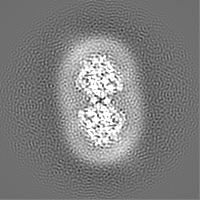
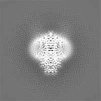
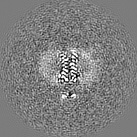
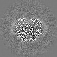
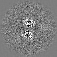
| Projections & slices | Image control
Images are generated by Spider. | ||||||||||||||||||||||||||||||||||||||||||||||||||||||||||||
| Voxel size | X=Y=Z: 1.07 Å | ||||||||||||||||||||||||||||||||||||||||||||||||||||||||||||
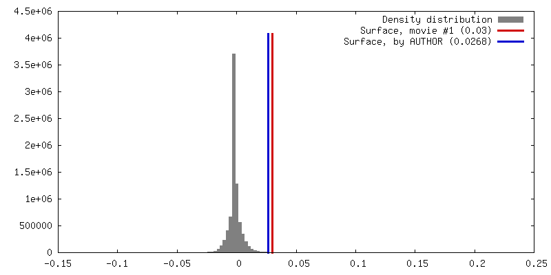
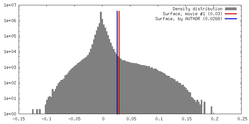
| Density |
| ||||||||||||||||||||||||||||||||||||||||||||||||||||||||||||
| Symmetry | Space group: 1 | ||||||||||||||||||||||||||||||||||||||||||||||||||||||||||||
| Details | EMDB XML:
CCP4 map header:
| ||||||||||||||||||||||||||||||||||||||||||||||||||||||||||||
 Z (Sec.)
 Y (Row.)
 X (Col.)


-Supplemental data
- Sample components
-Entire : Dimeric quinol dependent Nitric Oxide Reductase
| Entire | Name: Dimeric quinol dependent Nitric Oxide Reductase |
|---|---|
| Components |
|
-Supramolecule #1: Dimeric quinol dependent Nitric Oxide Reductase
| Supramolecule | Name: Dimeric quinol dependent Nitric Oxide Reductase / type: complex / ID: 1 / Parent: 0 / Macromolecule list: #1 |
|---|---|
| Source (natural) | Organism: Neisseria meningitidis alpha14 (bacteria) |
| Molecular weight | Theoretical: 150 KDa |
-Macromolecule #1: Nitric-oxide reductase
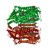
| Macromolecule | Name: Nitric-oxide reductase / type: protein_or_peptide / ID: 1 / Number of copies: 2 / Enantiomer: LEVO / EC number: nitric-oxide reductase |
|---|---|
| Source (natural) | Organism: Neisseria meningitidis alpha14 (bacteria) / Strain: alpha14 |
| Molecular weight | Theoretical: 84.389211 KDa |
| Recombinant expression | Organism: |
| Sequence | String: MGQYKKLWYL LFAVLAVCFT ILGYMGSEVY KKAPPYPEQV VSASGKVLMA KDDILAGQSA WQTTGGMEVG SVLGHGAYQA PDWTADWLH RELSAWLDLT AQQTYGKKFD EVSPEEQAVL KTRLADEYRN QSRIKEDGSV VISDTRVKAI ESILPYYHGV Y GDDPALQT ...String: MGQYKKLWYL LFAVLAVCFT ILGYMGSEVY KKAPPYPEQV VSASGKVLMA KDDILAGQSA WQTTGGMEVG SVLGHGAYQA PDWTADWLH RELSAWLDLT AQQTYGKKFD EVSPEEQAVL KTRLADEYRN QSRIKEDGSV VISDTRVKAI ESILPYYHGV Y GDDPALQT TREHFAMKNN TLPSQEAREK LFDFFFWTSW SASTNRPDET FTYTNNWPHE PLINNVPTTE NYMWSFTSVV LL LMGIGLL MWGYSFLTKH EEVEVPTEDP ISKVQLTPSQ KALGKYVFLT VALFVVQVLL GGLTAHYTVE GQGFYGIDEA LGF EMSDWF PYALTRTWHI QSAIFWIATG FLTAGLFLAP IVNGGKDPKF QRAGVNFLYI ALFIVVGGSY AGNFFALTHI LPPE FNFWF GHQGYEYLDL GRFWQLLLMV GLLLWLFLML RCTVSAFKEK GVDKNLLAIF VASMVGVGVF YAPGLFYGEK SPIAV MEYW RWWVVHLWVE GFFEVFATAA FAFVFYNMGF VRRSTATAST LAAAAIFMLG GVPGTLHHLY FSGSTSASMA IGACFS ALE VVPLVLLGRE AYEHWSYQHL SEWAKRLRWP LMCFVAVAFW NMIGAGVFGF LINPPISLFY IQGLNTSAVH AHAALFG VY GFLALGFVLL VARYLKPNVQ FDDKLMTWGF WLLNGGLVGM IAISLLPVGV IQAYASITHG LWYARSEEFL QMEILDTL R WVRTAADLIF IGGAICVAIQ ATKIVFGRDK UniProtKB: Nitric-oxide reductase |
-Macromolecule #2: PROTOPORPHYRIN IX CONTAINING FE
| Macromolecule | Name: PROTOPORPHYRIN IX CONTAINING FE / type: ligand / ID: 2 / Number of copies: 4 / Formula: HEM |
|---|---|
| Molecular weight | Theoretical: 616.487 Da |
| Chemical component information |  ChemComp-HEM: |
-Macromolecule #3: FE (III) ION
| Macromolecule | Name: FE (III) ION / type: ligand / ID: 3 / Number of copies: 2 / Formula: FE |
|---|---|
| Molecular weight | Theoretical: 55.845 Da |
-Macromolecule #4: CALCIUM ION
| Macromolecule | Name: CALCIUM ION / type: ligand / ID: 4 / Number of copies: 2 / Formula: CA |
|---|---|
| Molecular weight | Theoretical: 40.078 Da |
-Experimental details
-Structure determination
| Method | cryo EM |
|---|---|
 Processing Processing | single particle reconstruction |
| Aggregation state | particle |
-Sample preparation
| Concentration | 4.0 mg/mL |
|---|---|
| Buffer | pH: 8 |
| Grid | Model: Quantifoil R1.2/1.3 / Material: COPPER / Support film - Material: CARBON / Support film - topology: HOLEY / Pretreatment - Type: GLOW DISCHARGE |
| Vitrification | Cryogen name: ETHANE / Chamber humidity: 100 % / Chamber temperature: 277 K / Instrument: FEI VITROBOT MARK IV Details: Blot time of 6 seconds, with accompanying blot force of 6.. |
- Electron microscopy
| Microscope | FEI TITAN KRIOS |
|---|---|
| Image recording | Film or detector model: GATAN K2 SUMMIT (4k x 4k) / Detector mode: COUNTING / Digitization - Frames/image: 1-40 / Number grids imaged: 1 / Number real images: 3182 / Average exposure time: 8.0 sec. / Average electron dose: 69.44 e/Å2 |
| Electron beam | Acceleration voltage: 300 kV / Electron source:  FIELD EMISSION GUN FIELD EMISSION GUN |
| Electron optics | C2 aperture diameter: 70.0 µm / Illumination mode: FLOOD BEAM / Imaging mode: BRIGHT FIELD / Cs: 2.7 mm / Nominal defocus max: -3.5 µm / Nominal defocus min: -1.0 µm / Nominal magnification: 130000 |
| Sample stage | Specimen holder model: FEI TITAN KRIOS AUTOGRID HOLDER / Cooling holder cryogen: NITROGEN |
| Experimental equipment |  Model: Titan Krios / Image courtesy: FEI Company |