nitric-oxide reductase / cytochrome-c oxidase activity / aerobic respiration / oxidoreductase activity / heme binding / membrane / metal ion binding Similarity search - Function
: / Nitric oxide reductase subunit B, cytochrome c-like domain / Cytochrome C Oxidase; Chain A / Cytochrome c oxidase-like, subunit I domain / Cytochrome c oxidase subunit I / Cytochrome c oxidase-like, subunit I superfamily / Cytochrome C and Quinol oxidase polypeptide I / Up-down Bundle / Mainly Alpha Similarity search - Domain/homology
Biotechnology and Biological Sciences Research Council (BBSRC)
BB/L006960/1
United Kingdom
Biotechnology and Biological Sciences Research Council (BBSRC)
BB/N013972/1
United Kingdom
Citation
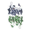
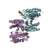
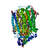
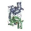
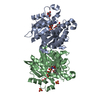
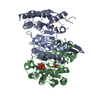
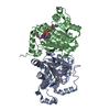
Journal: IUCrJ / Year: 2020 Title: The active form of quinol-dependent nitric oxide reductase from is a dimer. Authors: M Arif M Jamali / Chai C Gopalasingam / Rachel M Johnson / Takehiko Tosha / Kazumasa Muramoto / Stephen P Muench / Svetlana V Antonyuk / Yoshitsugu Shiro / Samar S Hasnain / Abstract: is carried by nearly a billion humans, causing developmental impairment and over 100 000 deaths a year. A quinol-dependent nitric oxide reductase (qNOR) plays a critical role in the survival of ... is carried by nearly a billion humans, causing developmental impairment and over 100 000 deaths a year. A quinol-dependent nitric oxide reductase (qNOR) plays a critical role in the survival of the bacterium in the human host. X-ray crystallographic analyses of qNOR, including that from (qNOR) reported here at 3.15 Å resolution, show monomeric assemblies, despite the more active dimeric sample being used for crystallization. Cryo-electron microscopic analysis of the same chromatographic fraction of qNOR, however, revealed a dimeric assembly at 3.06 Å resolution. It is shown that zinc (which is used in crystallization) binding near the dimer-stabilizing TMII region contributes to the disruption of the dimer. A similar destabilization is observed in the monomeric (∼85 kDa) cryo-EM structure of a mutant (Glu494Ala) qNOR from the opportunistic pathogen () , which primarily migrates as a monomer. The monomer-dimer transition of qNORs seen in the cryo-EM and crystallographic structures has wider implications for structural studies of multimeric membrane proteins. X-ray crystallographic and cryo-EM structural analyses have been performed on the same chromatographic fraction of qNOR to high resolution. This represents one of the first examples in which the two approaches have been used to reveal a monomeric assembly and a dimeric assembly in vitrified cryo-EM grids. A number of factors have been identified that may trigger the destabilization of helices that are necessary to preserve the integrity of the dimer. These include zinc binding near the entry of the putative proton-transfer channel and the preservation of the conformational integrity of the active site. The mutation near the active site results in disruption of the active site, causing an additional destabilization of helices (TMIX and TMX) that flank the proton-transfer channel helices, creating an inert monomeric enzyme.
Method to determine structure: MIR / Resolution: 3.15→49.92 Å / Cor.coef. Fo:Fc: 0.907 / Cor.coef. Fo:Fc free: 0.903 / SU B: 31.933 / SU ML: 0.498 / Cross valid method: THROUGHOUT / ESU R Free: 0.566 / Stereochemistry target values: MAXIMUM LIKELIHOOD Details: SF FILE CONTAINS FRIEDEL PAIRS UNDER I/F_MINUS AND I/F_PLUS COLUMNS.
Rfactor
Num. reflection
% reflection
Selection details
Rfree
0.30253
1063
5.3 %
RANDOM
Rwork
0.25029
-
-
-
obs
0.25313
19034
81.46 %
-
Solvent computation
Ion probe radii: 0.8 Å / Shrinkage radii: 0.8 Å / VDW probe radii: 1.2 Å / Solvent model: MASK
 Movie
Movie Controller

 Yorodumi
 Open data
 Basic information
 Components
 Keywords
 Function and homology information
 Neisseria meningitidis (bacteria)
 X-RAY DIFFRACTION /
  Authors
 Japan,
  United Kingdom, 5items
  Citation
 Structure visualization
 Downloads & links
 Other downloads
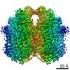


 PDBj
PDBj

 Assembly


 Mass: 616.487 Da / Num. of mol.: 2 / Source method: obtained synthetically / Formula: C34H32FeN4O4
 Mass: 55.845 Da / Num. of mol.: 1 / Source method: obtained synthetically / Formula: Fe / Feature type: SUBJECT OF INVESTIGATION
 Mass: 40.078 Da / Num. of mol.: 1 / Source method: obtained synthetically / Formula: Ca
 Mass: 65.409 Da / Num. of mol.: 3 / Source method: obtained synthetically / Formula: Zn
 Sample preparation
 Processing