 Movie
Movie Controller


+ Open data

- Basic information
| Entry | Database: PDB / ID: 6fwf | |||||||||
|---|---|---|---|---|---|---|---|---|---|---|
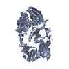
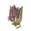
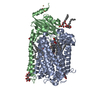
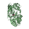
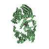
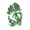
| Title | Low resolution structure of Neisseria meningitidis qNOR | |||||||||
 Components Components | Nitric-oxide reductase | |||||||||
 Keywords Keywords | OXIDOREDUCTASE / reductase / membrane-bound / nitric oxide | |||||||||
| Function / homology |  Function and homology information Function and homology informationnitric-oxide reductase / cytochrome-c oxidase activity / aerobic respiration / oxidoreductase activity / heme binding / membrane / metal ion binding Similarity search - Function | |||||||||
| Biological species |  Neisseria meningitidis (bacteria) Neisseria meningitidis (bacteria) | |||||||||
| Method |  X-RAY DIFFRACTION / SYNCHROTRON / MOLECULAR REPLACEMENT / Resolution: 4.2 Å X-RAY DIFFRACTION / SYNCHROTRON / MOLECULAR REPLACEMENT / Resolution: 4.2 Å | |||||||||
 Authors Authors | Young, D. / Antonyuk, S. / Tosha, T. / Hisano, T. / Hasnain, S. / Shiro, Y. | |||||||||
 Citation Citation | Journal: Sci Rep / Year: 2018 Title: Characterization of the quinol-dependent nitric oxide reductase from the pathogen Neisseria meningitidis, an electrogenic enzyme. Authors: Gonska, N. / Young, D. / Yuki, R. / Okamoto, T. / Hisano, T. / Antonyuk, S. / Hasnain, S.S. / Muramoto, K. / Shiro, Y. / Tosha, T. / Adelroth, P. | |||||||||
| History |
|
- Structure visualization
| Structure viewer | Molecule: MolmilJmol/JSmol |
|---|
- Downloads & links
-Download
| PDBx/mmCIF format | 6fwf.cif.gz | 160.2 KB | Display | PDBx/mmCIF format |
|---|---|---|---|---|
| PDB format | pdb6fwf.ent.gz | 124.9 KB | Display | PDB format |
| PDBx/mmJSON format | 6fwf.json.gz | Tree view | PDBx/mmJSON format | |
| Others |  Other downloads Other downloads |
-Validation report
| Arichive directory | https://data.pdbj.org/pub/pdb/validation_reports/fw/6fwfftp://data.pdbj.org/pub/pdb/validation_reports/fw/6fwf | HTTPS FTP |
|---|
-Related structure data
| Related structure data | 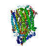 3ayfS S: Starting model for refinement |
|---|---|
| Similar structure data |


-Links
 PDBj
PDBj

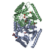
- Assembly
| Deposited unit | 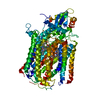
| ||||||||
|---|---|---|---|---|---|---|---|---|---|
| 1 |
| ||||||||
| Unit cell |
|
-Components
| #1: Protein | Mass: 84389.211 Da / Num. of mol.: 1 Source method: isolated from a genetically manipulated source Source: (gene. exp.) Neisseria meningitidis (strain alpha14) (bacteria)Strain: alpha14 / Gene: norB, NMO_1451 / Plasmid: pRSET-C / Production host: | ||||
|---|---|---|---|---|---|
| #2: Chemical |   Mass: 616.487 Da / Num. of mol.: 2 / Source method: obtained synthetically / Formula: C34H32FeN4O4 Mass: 616.487 Da / Num. of mol.: 2 / Source method: obtained synthetically / Formula: C34H32FeN4O4#3: Chemical | ChemComp-CA / |   Mass: 40.078 Da / Num. of mol.: 1 / Source method: obtained synthetically / Formula: Ca Mass: 40.078 Da / Num. of mol.: 1 / Source method: obtained synthetically / Formula: Ca#4: Chemical | ChemComp-FE / |   Mass: 55.845 Da / Num. of mol.: 1 / Source method: obtained synthetically / Formula: Fe Mass: 55.845 Da / Num. of mol.: 1 / Source method: obtained synthetically / Formula: Fe |
-Experimental details
-Experiment
| Experiment | Method: X-RAY DIFFRACTION / Number of used crystals: 1 |
|---|
- Sample preparation
| Crystal | Density Matthews: 4.47 Å3/Da / Density % sol: 72.48 % |
|---|---|
| Crystal grow | Temperature: 283.15 K / Method: vapor diffusion, sitting drop / pH: 6.5 Details: PEG 400, cadmium chloride, magnesium chloride, 2-(N-morpholino)ethanesulfonic acid |
-Data collection
| Diffraction | Mean temperature: 100 K | ||||||||||||||||||||||||
|---|---|---|---|---|---|---|---|---|---|---|---|---|---|---|---|---|---|---|---|---|---|---|---|---|---|
| Diffraction source | Source: SYNCHROTRON / Site: SPring-8  / Beamline: BL41XU / Wavelength: 1 Å / Beamline: BL41XU / Wavelength: 1 Å | ||||||||||||||||||||||||
| Detector | Type: RAYONIX MX225HE / Detector: CCD / Date: Dec 11, 2013 | ||||||||||||||||||||||||
| Radiation | Protocol: SINGLE WAVELENGTH / Monochromatic (M) / Laue (L): M / Scattering type: x-ray | ||||||||||||||||||||||||
| Radiation wavelength | Wavelength: 1 Å / Relative weight: 1 | ||||||||||||||||||||||||
| Reflection | Resolution: 4.2→53.64 Å / Num. obs: 11511 / % possible obs: 99.86 % / Redundancy: 14.4 % / Biso Wilson estimate: 170.75 Å2 / CC1/2: 0.998 / Rmerge(I) obs: 0.251 / Rpim(I) all: 0.07 / Rrim(I) all: 0.261 / Net I/σ(I): 5.5 | ||||||||||||||||||||||||
| Reflection shell | Diffraction-ID: 1
|
- Processing
| Software |
| ||||||||||||||||||||||||||||||||||||||||||||||||||||||||||||
|---|---|---|---|---|---|---|---|---|---|---|---|---|---|---|---|---|---|---|---|---|---|---|---|---|---|---|---|---|---|---|---|---|---|---|---|---|---|---|---|---|---|---|---|---|---|---|---|---|---|---|---|---|---|---|---|---|---|---|---|---|---|
| Refinement | Method to determine structure: MOLECULAR REPLACEMENT Starting model: 3AYF Resolution: 4.2→53.64 Å / Cor.coef. Fo:Fc: 0.85 / Cor.coef. Fo:Fc free: 0.82 / SU B: 137.639 / SU ML: 1.627 / Cross valid method: THROUGHOUT / σ(F): 0 / ESU R Free: 1.153 Details: HYDROGENS HAVE BEEN ADDED IN THE RIDING POSITIONS U VALUES : REFINED INDIVIDUALLY
| ||||||||||||||||||||||||||||||||||||||||||||||||||||||||||||
| Solvent computation | Ion probe radii: 0.8 Å / Shrinkage radii: 0.8 Å / VDW probe radii: 1.2 Å | ||||||||||||||||||||||||||||||||||||||||||||||||||||||||||||
| Displacement parameters | Biso max: 616.17 Å2 / Biso mean: 287.437 Å2 / Biso min: 99.6 Å2
| ||||||||||||||||||||||||||||||||||||||||||||||||||||||||||||
| Refinement step | Cycle: final / Resolution: 4.2→53.64 Å
| ||||||||||||||||||||||||||||||||||||||||||||||||||||||||||||
| Refine LS restraints |
| ||||||||||||||||||||||||||||||||||||||||||||||||||||||||||||
| LS refinement shell | Resolution: 4.2→4.309 Å / Rfactor Rfree error: 0 / Total num. of bins used: 20
|
