 Movie
Movie Controller


+ Open data

- Basic information
| Entry | Database: PDB / ID: 6knk | |||||||||
|---|---|---|---|---|---|---|---|---|---|---|
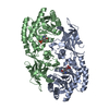
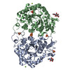
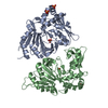
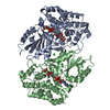
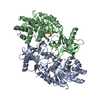
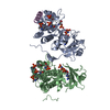
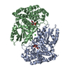
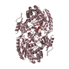
| Title | Crystal structure of SbnH in complex with citryl-diaminoethane | |||||||||
 Components Components | Probable diaminopimelate decarboxylase protein | |||||||||
 Keywords Keywords | BIOSYNTHETIC PROTEIN / PLP-dependent decarboxylase / Staphyloferrin B | |||||||||
| Function / homology |  Function and homology information Function and homology informationdiaminopimelate decarboxylase activity / polyamine biosynthetic process / : Similarity search - Function | |||||||||
| Biological species |  Staphylococcus aureus subsp. aureus Mu50 (bacteria) Staphylococcus aureus subsp. aureus Mu50 (bacteria) | |||||||||
| Method |  X-RAY DIFFRACTION / SYNCHROTRON / MOLECULAR REPLACEMENT / Resolution: 2.3 Å X-RAY DIFFRACTION / SYNCHROTRON / MOLECULAR REPLACEMENT / Resolution: 2.3 Å | |||||||||
 Authors Authors | Tang, J. / Ju, Y. / Zhou, H. | |||||||||
| Funding support |  China, 2items China, 2items
| |||||||||
 Citation Citation | Journal: J.Mol.Biol. / Year: 2019 Title: Structural Insights into Substrate Recognition and Activity Regulation of the Key Decarboxylase SbnH in Staphyloferrin B Biosynthesis. Authors: Tang, J. / Ju, Y. / Gu, Q. / Xu, J. / Zhou, H. | |||||||||
| History |
|
- Structure visualization
| Structure viewer | Molecule: MolmilJmol/JSmol |
|---|
- Downloads & links
-Download
| PDBx/mmCIF format | 6knk.cif.gz | 243.1 KB | Display | PDBx/mmCIF format |
|---|---|---|---|---|
| PDB format | pdb6knk.ent.gz | 192.6 KB | Display | PDB format |
| PDBx/mmJSON format | 6knk.json.gz | Tree view | PDBx/mmJSON format | |
| Others |  Other downloads Other downloads |
-Validation report
| Arichive directory | https://data.pdbj.org/pub/pdb/validation_reports/kn/6knkftp://data.pdbj.org/pub/pdb/validation_reports/kn/6knk | HTTPS FTP |
|---|
-Related structure data
| Related structure data | 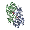 6knhC  6kniC  2j66S S: Starting model for refinement C: citing same article ( |
|---|---|
| Similar structure data |


-Links
 PDBj
PDBj
- Assembly
| Deposited unit | 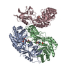
| ||||||||
|---|---|---|---|---|---|---|---|---|---|
| 1 | 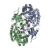
| ||||||||
| 2 | 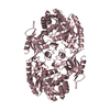
| ||||||||
| Unit cell |
|
-Components
| #1: Protein | Mass: 46536.312 Da / Num. of mol.: 3 / Mutation: K50A Source method: isolated from a genetically manipulated source Source: (gene. exp.) Staphylococcus aureus subsp. aureus Mu50 (bacteria)Strain: Mu50 / Gene: SAV0123 / Production host: #2: Chemical | ChemComp-DN9 / ( |   Mass: 465.349 Da / Num. of mol.: 1 / Source method: obtained synthetically / Formula: C16H24N3O11P / Feature type: SUBJECT OF INVESTIGATION Mass: 465.349 Da / Num. of mol.: 1 / Source method: obtained synthetically / Formula: C16H24N3O11P / Feature type: SUBJECT OF INVESTIGATION#3: Chemical |   Mass: 94.971 Da / Num. of mol.: 2 / Source method: obtained synthetically / Formula: PO4 Mass: 94.971 Da / Num. of mol.: 2 / Source method: obtained synthetically / Formula: PO4#4: Chemical | ChemComp-X3J / ( |   Mass: 234.207 Da / Num. of mol.: 1 / Source method: obtained synthetically / Formula: C8H14N2O6 / Feature type: SUBJECT OF INVESTIGATION Mass: 234.207 Da / Num. of mol.: 1 / Source method: obtained synthetically / Formula: C8H14N2O6 / Feature type: SUBJECT OF INVESTIGATION#5: Water | ChemComp-HOH / |  Mass: 18.015 Da / Num. of mol.: 274 / Source method: isolated from a natural source / Formula: H2O Mass: 18.015 Da / Num. of mol.: 274 / Source method: isolated from a natural source / Formula: H2OHas ligand of interest | Y | |
|---|
-Experimental details
-Experiment
| Experiment | Method: X-RAY DIFFRACTION / Number of used crystals: 1 |
|---|
- Sample preparation
| Crystal | Density Matthews: 2.94 Å3/Da / Density % sol: 58.14 % |
|---|---|
| Crystal grow | Temperature: 290 K / Method: evaporation Details: 0.2 M Na Citrate, 50 mM Bis-Tris pH 6.0, 23% PEG 3,350 |
-Data collection
| Diffraction | Mean temperature: 100 K / Serial crystal experiment: N |
|---|---|
| Diffraction source | Source: SYNCHROTRON / Site: SSRF / Beamline: BL19U1 / Wavelength: 0.9791 Å |
| Detector | Type: DECTRIS PILATUS3 S 6M / Detector: PIXEL / Date: Dec 16, 2018 |
| Radiation | Protocol: SINGLE WAVELENGTH / Monochromatic (M) / Laue (L): M / Scattering type: x-ray |
| Radiation wavelength | Wavelength: 0.9791 Å / Relative weight: 1 |
| Reflection | Resolution: 2.3→46.74 Å / Num. obs: 65898 / % possible obs: 97.3 % / Redundancy: 3.3 % / Rmerge(I) obs: 0.047 / Net I/σ(I): 15.1 |
| Reflection shell | Resolution: 2.3→2.35 Å / Rmerge(I) obs: 0.367 / Num. unique obs: 4430 |
- Processing
| Software |
| ||||||||||||||||||||||||||||||||||||||||||||||||||||||||||||||||||||||||||||||||||||||||||||||||||||||||||||||||||||||||||||||||||||||||||||||||||||||||||||||||||||||||||||||||||||||
|---|---|---|---|---|---|---|---|---|---|---|---|---|---|---|---|---|---|---|---|---|---|---|---|---|---|---|---|---|---|---|---|---|---|---|---|---|---|---|---|---|---|---|---|---|---|---|---|---|---|---|---|---|---|---|---|---|---|---|---|---|---|---|---|---|---|---|---|---|---|---|---|---|---|---|---|---|---|---|---|---|---|---|---|---|---|---|---|---|---|---|---|---|---|---|---|---|---|---|---|---|---|---|---|---|---|---|---|---|---|---|---|---|---|---|---|---|---|---|---|---|---|---|---|---|---|---|---|---|---|---|---|---|---|---|---|---|---|---|---|---|---|---|---|---|---|---|---|---|---|---|---|---|---|---|---|---|---|---|---|---|---|---|---|---|---|---|---|---|---|---|---|---|---|---|---|---|---|---|---|---|---|---|---|
| Refinement | Method to determine structure: MOLECULAR REPLACEMENT Starting model: 2J66 Resolution: 2.3→46.74 Å / Cor.coef. Fo:Fc: 0.941 / Cor.coef. Fo:Fc free: 0.932 / SU B: 9.466 / SU ML: 0.211 / Cross valid method: THROUGHOUT / σ(F): 0 / ESU R: 0.327 / ESU R Free: 0.227 / Stereochemistry target values: MAXIMUM LIKELIHOOD Details: HYDROGENS HAVE BEEN ADDED IN THE RIDING POSITIONS U VALUES : REFINED INDIVIDUALL
| ||||||||||||||||||||||||||||||||||||||||||||||||||||||||||||||||||||||||||||||||||||||||||||||||||||||||||||||||||||||||||||||||||||||||||||||||||||||||||||||||||||||||||||||||||||||
| Solvent computation | Ion probe radii: 0.8 Å / Shrinkage radii: 0.8 Å / VDW probe radii: 1.2 Å / Solvent model: MASK | ||||||||||||||||||||||||||||||||||||||||||||||||||||||||||||||||||||||||||||||||||||||||||||||||||||||||||||||||||||||||||||||||||||||||||||||||||||||||||||||||||||||||||||||||||||||
| Displacement parameters | Biso max: 75.6 Å2 / Biso mean: 41.074 Å2 / Biso min: 22.1 Å2
| ||||||||||||||||||||||||||||||||||||||||||||||||||||||||||||||||||||||||||||||||||||||||||||||||||||||||||||||||||||||||||||||||||||||||||||||||||||||||||||||||||||||||||||||||||||||
| Refinement step | Cycle: final / Resolution: 2.3→46.74 Å
| ||||||||||||||||||||||||||||||||||||||||||||||||||||||||||||||||||||||||||||||||||||||||||||||||||||||||||||||||||||||||||||||||||||||||||||||||||||||||||||||||||||||||||||||||||||||
| Refine LS restraints |
|