 Movie
Movie Controller

[English] 日本語
 Yorodumi
- PDB-6kni: Crystal structure of SbnH in complex with the cofactor PLP, a PLP... -
+ Open data

- Basic information
| Entry | Database: PDB / ID: 6kni | |||||||||
|---|---|---|---|---|---|---|---|---|---|---|
| Title | Crystal structure of SbnH in complex with the cofactor PLP, a PLP-dependent decarboxylase in Staphyloferrin B biothesynthesis | |||||||||
 Components Components | Probable diaminopimelate decarboxylase protein | |||||||||
 Keywords Keywords | BIOSYNTHETIC PROTEIN / PLP-dependent decarboxylase / Staphyloferrin B | |||||||||
| Function / homology |  Function and homology information Function and homology informationdiaminopimelate decarboxylase activity / polyamine biosynthetic process / : Similarity search - Function | |||||||||
| Biological species |  Staphylococcus aureus subsp. aureus Mu50 (bacteria) Staphylococcus aureus subsp. aureus Mu50 (bacteria) | |||||||||
| Method |  X-RAY DIFFRACTION / SYNCHROTRON / MOLECULAR REPLACEMENT / Resolution: 1.97 Å X-RAY DIFFRACTION / SYNCHROTRON / MOLECULAR REPLACEMENT / Resolution: 1.97 Å | |||||||||
 Authors Authors | Tang, J. / Ju, Y. / Zhou, H. | |||||||||
| Funding support |  China, 2items China, 2items
| |||||||||
 Citation Citation | Journal: J.Mol.Biol. / Year: 2019 Title: Structural Insights into Substrate Recognition and Activity Regulation of the Key Decarboxylase SbnH in Staphyloferrin B Biosynthesis. Authors: Tang, J. / Ju, Y. / Gu, Q. / Xu, J. / Zhou, H. | |||||||||
| History |
|
- Structure visualization
| Structure viewer | Molecule: MolmilJmol/JSmol |
|---|
- Downloads & links
-Download
| PDBx/mmCIF format | 6kni.cif.gz | 249.3 KB | Display | PDBx/mmCIF format |
|---|---|---|---|---|
| PDB format | pdb6kni.ent.gz | 198.9 KB | Display | PDB format |
| PDBx/mmJSON format | 6kni.json.gz | Tree view | PDBx/mmJSON format | |
| Others |  Other downloads Other downloads |
-Validation report
| Arichive directory | https://data.pdbj.org/pub/pdb/validation_reports/kn/6kniftp://data.pdbj.org/pub/pdb/validation_reports/kn/6kni | HTTPS FTP |
|---|
-Related structure data
| Related structure data |  6knhC  6knkC  2j66S S: Starting model for refinement C: citing same article ( |
|---|---|
| Similar structure data |


-Links
 PDBj
PDBj
- Assembly
| Deposited unit | 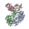
| ||||||||
|---|---|---|---|---|---|---|---|---|---|
| 1 | 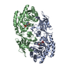
| ||||||||
| 2 | 
| ||||||||
| Unit cell |
|
-Components
| #1: Protein | Mass: 46594.414 Da / Num. of mol.: 3 Source method: isolated from a genetically manipulated source Source: (gene. exp.) Staphylococcus aureus subsp. aureus Mu50 (bacteria)Strain: Mu50 / Gene: SAV0123 / Production host: #2: Chemical |   Mass: 247.142 Da / Num. of mol.: 3 / Source method: obtained synthetically / Formula: C8H10NO6P Mass: 247.142 Da / Num. of mol.: 3 / Source method: obtained synthetically / Formula: C8H10NO6P#3: Water | ChemComp-HOH / |  Mass: 18.015 Da / Num. of mol.: 591 / Source method: isolated from a natural source / Formula: H2O Mass: 18.015 Da / Num. of mol.: 591 / Source method: isolated from a natural source / Formula: H2OHas ligand of interest | N | |
|---|
-Experimental details
-Experiment
| Experiment | Method: X-RAY DIFFRACTION / Number of used crystals: 1 |
|---|
- Sample preparation
| Crystal | Density Matthews: 2.96 Å3/Da / Density % sol: 58.39 % |
|---|---|
| Crystal grow | Temperature: 290 K / Method: evaporation / Details: 0.2 M NaAC, 50 mM Bis-Tris pH 6.0, 23% PEG 3,350 |
-Data collection
| Diffraction | Mean temperature: 100 K / Serial crystal experiment: N |
|---|---|
| Diffraction source | Source: SYNCHROTRON / Site: SSRF / Beamline: BL17U1 / Wavelength: 0.979 Å |
| Detector | Type: ADSC QUANTUM 315r / Detector: CCD / Date: Dec 28, 2017 |
| Radiation | Protocol: SINGLE WAVELENGTH / Monochromatic (M) / Laue (L): M / Scattering type: x-ray |
| Radiation wavelength | Wavelength: 0.979 Å / Relative weight: 1 |
| Reflection | Resolution: 1.97→109.32 Å / Num. obs: 105438 / % possible obs: 97.7 % / Redundancy: 3.4 % / Rmerge(I) obs: 0.097 / Net I/σ(I): 9.1 |
| Reflection shell | Resolution: 1.97→2.08 Å / Rmerge(I) obs: 0.443 / Num. unique obs: 15457 |
- Processing
| Software |
| ||||||||||||||||
|---|---|---|---|---|---|---|---|---|---|---|---|---|---|---|---|---|---|
| Refinement | Method to determine structure: MOLECULAR REPLACEMENT Starting model: 2j66 Resolution: 1.97→50 Å / Cross valid method: FREE R-VALUE
| ||||||||||||||||
| Refinement step | Cycle: LAST / Resolution: 1.97→50 Å
| ||||||||||||||||
| LS refinement shell | Resolution: 1.97→2.021 Å
|
