 Movie
Movie Controller

[English] 日本語
 Yorodumi
- PDB-6qjo: DNA containing both right- and left-handed parallel-stranded G-qu... -
+ Open data

- Basic information
| Entry | Database: PDB / ID: 6qjo | ||||||
|---|---|---|---|---|---|---|---|
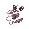
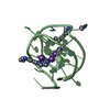
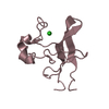
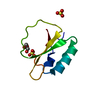
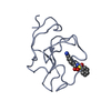
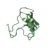
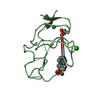
| Title | DNA containing both right- and left-handed parallel-stranded G-quadruplexes | ||||||
 Components Components | DNA (28-MER) | ||||||
 Keywords Keywords | DNA / G-quadruplex left-handed right-handed | ||||||
| Function / homology | : / DNA / DNA (> 10) Function and homology information Function and homology information | ||||||
| Biological species | synthetic construct (others) | ||||||
| Method |  X-RAY DIFFRACTION / SYNCHROTRON / MOLECULAR REPLACEMENT / Resolution: 1.8 Å X-RAY DIFFRACTION / SYNCHROTRON / MOLECULAR REPLACEMENT / Resolution: 1.8 Å | ||||||
 Authors Authors | Winnerdy, F.R. / Bakalar, B. / Maity, A. / Vandana, J.J. / Schmitt, E. / Mechulam, Y. / Phan, A.T. | ||||||
 Citation Citation | Journal: Nucleic Acids Res. / Year: 2019 Title: NMR solution and X-ray crystal structures of a DNA molecule containing both right- and left-handed parallel-stranded G-quadruplexes. Authors: Winnerdy, F.R. / Bakalar, B. / Maity, A. / Vandana, J.J. / Mechulam, Y. / Schmitt, E. / Phan, A.T. | ||||||
| History |
|
- Structure visualization
| Structure viewer | Molecule: MolmilJmol/JSmol |
|---|
- Downloads & links
-Download
| PDBx/mmCIF format | 6qjo.cif.gz | 136.7 KB | Display | PDBx/mmCIF format |
|---|---|---|---|---|
| PDB format | pdb6qjo.ent.gz | 107 KB | Display | PDB format |
| PDBx/mmJSON format | 6qjo.json.gz | Tree view | PDBx/mmJSON format | |
| Others |  Other downloads Other downloads |
-Validation report
| Arichive directory | https://data.pdbj.org/pub/pdb/validation_reports/qj/6qjoftp://data.pdbj.org/pub/pdb/validation_reports/qj/6qjo | HTTPS FTP |
|---|
-Related structure data
| Related structure data |  6jceC 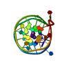 4u5mS S: Starting model for refinement C: citing same article ( |
|---|---|
| Similar structure data |


-Links
 PDBj
PDBj


- Assembly
| Deposited unit | 
| ||||||||
|---|---|---|---|---|---|---|---|---|---|
| 1 | 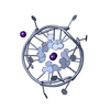
| ||||||||
| 2 | 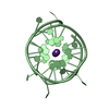
| ||||||||
| 3 | 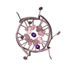
| ||||||||
| 4 | 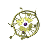
| ||||||||
| Unit cell |
|
-Components
| #1: DNA chain | Mass: 8897.662 Da / Num. of mol.: 4 Source method: isolated from a genetically manipulated source Source: (gene. exp.) synthetic construct (others) / Production host: synthetic construct (others) #2: Chemical | ChemComp-K /   Mass: 39.098 Da / Num. of mol.: 15 / Source method: obtained synthetically / Formula: K Mass: 39.098 Da / Num. of mol.: 15 / Source method: obtained synthetically / Formula: K#3: Water | ChemComp-HOH / |  Mass: 18.015 Da / Num. of mol.: 166 / Source method: isolated from a natural source / Formula: H2O Mass: 18.015 Da / Num. of mol.: 166 / Source method: isolated from a natural source / Formula: H2O |
|---|
-Experimental details
-Experiment
| Experiment | Method: X-RAY DIFFRACTION / Number of used crystals: 1 |
|---|
- Sample preparation
| Crystal | Density Matthews: 1.82 Å3/Da / Density % sol: 32.54 % |
|---|---|
| Crystal grow | Temperature: 297 K / Method: vapor diffusion, sitting drop / pH: 7 / Details: 60% MPD |
-Data collection
| Diffraction | Mean temperature: 100 K / Serial crystal experiment: N |
|---|---|
| Diffraction source | Source: SYNCHROTRON / Site: SOLEIL  / Beamline: PROXIMA 1 / Wavelength: 0.98 Å / Beamline: PROXIMA 1 / Wavelength: 0.98 Å |
| Detector | Type: DECTRIS PILATUS 6M-F / Detector: PIXEL / Date: Jan 28, 2018 |
| Radiation | Protocol: SINGLE WAVELENGTH / Monochromatic (M) / Laue (L): M / Scattering type: x-ray |
| Radiation wavelength | Wavelength: 0.98 Å / Relative weight: 1 |
| Reflection | Resolution: 1.8→50 Å / Num. obs: 22501 / % possible obs: 95.9 % / Redundancy: 4.1 % / Biso Wilson estimate: 25.3 Å2 / CC1/2: 0.99 / Rsym value: 0.115 / Net I/σ(I): 10.45 |
| Reflection shell | Resolution: 1.8→1.9 Å / Redundancy: 2.72 % / Mean I/σ(I) obs: 5.8 / Num. unique obs: 3079 / CC1/2: 0.818 / Rsym value: 0.359 / % possible all: 88 |
- Processing
| Software |
| |||||||||||||||||||||||||||||||||||||||||||||||||||||||||||||||
|---|---|---|---|---|---|---|---|---|---|---|---|---|---|---|---|---|---|---|---|---|---|---|---|---|---|---|---|---|---|---|---|---|---|---|---|---|---|---|---|---|---|---|---|---|---|---|---|---|---|---|---|---|---|---|---|---|---|---|---|---|---|---|---|---|
| Refinement | Method to determine structure: MOLECULAR REPLACEMENT Starting model: 4U5M Resolution: 1.8→39.27 Å / Cross valid method: FREE R-VALUE / σ(F): 2.77 / Phase error: 24.09
| |||||||||||||||||||||||||||||||||||||||||||||||||||||||||||||||
| Solvent computation | Shrinkage radii: 0.9 Å / VDW probe radii: 1.11 Å | |||||||||||||||||||||||||||||||||||||||||||||||||||||||||||||||
| Refinement step | Cycle: LAST / Resolution: 1.8→39.27 Å
| |||||||||||||||||||||||||||||||||||||||||||||||||||||||||||||||
| Refine LS restraints |
| |||||||||||||||||||||||||||||||||||||||||||||||||||||||||||||||
| LS refinement shell |
|
