 Movie
Movie Controller


+ Open data

- Basic information
| Entry | Database: PDB / ID: 7d5e | ||||||
|---|---|---|---|---|---|---|---|
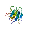
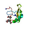
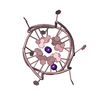
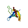
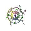
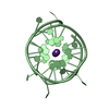
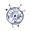
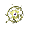
| Title | Left-handed G-quadruplex containing two bulges | ||||||
 Components Components | 2xBulge-LHG4motif | ||||||
 Keywords Keywords | DNA / G-quadruplex / Bulge / Left-handed | ||||||
| Function / homology | : / SPERMINE / DNA / DNA (> 10) Function and homology information Function and homology information | ||||||
| Biological species | synthetic construct (others) | ||||||
| Method |  X-RAY DIFFRACTION / SYNCHROTRON / MOLECULAR REPLACEMENT / Resolution: 1.296 Å X-RAY DIFFRACTION / SYNCHROTRON / MOLECULAR REPLACEMENT / Resolution: 1.296 Å | ||||||
 Authors Authors | Das, P. / Maity, A. / Ngo, K.H. / Winnerdy, F.R. / Bakalar, B. / Mechulam, Y. / Schmitt, E. / Phan, A.T. | ||||||
| Funding support |  Singapore, 1items Singapore, 1items
| ||||||
 Citation Citation | Journal: Nucleic Acids Res. / Year: 2021 Title: Bulges in left-handed G-quadruplexes. Authors: Das, P. / Ngo, K.H. / Winnerdy, F.R. / Maity, A. / Bakalar, B. / Mechulam, Y. / Schmitt, E. / Phan, A.T. | ||||||
| History |
|
- Structure visualization
| Structure viewer | Molecule: MolmilJmol/JSmol |
|---|
- Downloads & links
-Download
| PDBx/mmCIF format | 7d5e.cif.gz | 77.4 KB | Display | PDBx/mmCIF format |
|---|---|---|---|---|
| PDB format | pdb7d5e.ent.gz | 58.1 KB | Display | PDB format |
| PDBx/mmJSON format | 7d5e.json.gz | Tree view | PDBx/mmJSON format | |
| Others |  Other downloads Other downloads |
-Validation report
| Arichive directory | https://data.pdbj.org/pub/pdb/validation_reports/d5/7d5eftp://data.pdbj.org/pub/pdb/validation_reports/d5/7d5e | HTTPS FTP |
|---|
-Related structure data
| Related structure data | 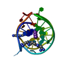 7d5dC 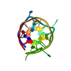 7d5fC 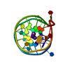 4u5mS S: Starting model for refinement C: citing same article ( |
|---|---|
| Similar structure data |

-Links
 PDBj
PDBj


- Assembly
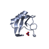
| Deposited unit | 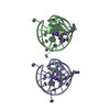
| ||||||||
|---|---|---|---|---|---|---|---|---|---|
| 1 | 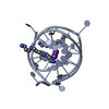
| ||||||||
| 2 | 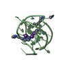
| ||||||||
| Unit cell |
|
-Components
| #1: DNA chain | Mass: 8872.649 Da / Num. of mol.: 2 / Source method: obtained synthetically / Source: (synth.) synthetic construct (others) #2: Chemical | ChemComp-K /   Mass: 39.098 Da / Num. of mol.: 7 / Source method: obtained synthetically / Formula: K Mass: 39.098 Da / Num. of mol.: 7 / Source method: obtained synthetically / Formula: K#3: Chemical | ChemComp-SPM /   Mass: 202.340 Da / Num. of mol.: 4 / Source method: obtained synthetically / Formula: C10H26N4 Mass: 202.340 Da / Num. of mol.: 4 / Source method: obtained synthetically / Formula: C10H26N4#4: Chemical | ChemComp-NA / |   Mass: 22.990 Da / Num. of mol.: 1 / Source method: obtained synthetically / Formula: Na Mass: 22.990 Da / Num. of mol.: 1 / Source method: obtained synthetically / Formula: Na#5: Water | ChemComp-HOH / |  Mass: 18.015 Da / Num. of mol.: 208 / Source method: isolated from a natural source / Formula: H2O Mass: 18.015 Da / Num. of mol.: 208 / Source method: isolated from a natural source / Formula: H2OHas ligand of interest | N | |
|---|
-Experimental details
-Experiment
| Experiment | Method: X-RAY DIFFRACTION / Number of used crystals: 1 |
|---|
- Sample preparation
| Crystal | Density Matthews: 1.79 Å3/Da / Density % sol: 31.43 % |
|---|---|
| Crystal grow | Temperature: 297 K / Method: vapor diffusion, sitting drop / pH: 7 Details: 0.08 M potassium chloride, 0.04 M sodium cacodylate trihydrate pH 7.0, 60% v/v -2-methyl-2,4-pentanediol and 0.012 M spermine tetrahydrochloride |
-Data collection
| Diffraction | Mean temperature: 100 K / Serial crystal experiment: N |
|---|---|
| Diffraction source | Source: SYNCHROTRON / Site: SOLEIL  / Beamline: PROXIMA 1 / Wavelength: 0.9793 Å / Beamline: PROXIMA 1 / Wavelength: 0.9793 Å |
| Detector | Type: DECTRIS PILATUS 6M / Detector: PIXEL / Date: Jun 30, 2017 |
| Radiation | Protocol: SINGLE WAVELENGTH / Monochromatic (M) / Laue (L): M / Scattering type: x-ray |
| Radiation wavelength | Wavelength: 0.9793 Å / Relative weight: 1 |
| Reflection | Resolution: 1.296→29.093 Å / Num. obs: 31051 / % possible obs: 94.67 % / Redundancy: 6 % / Biso Wilson estimate: 11.81 Å2 / CC1/2: 0.996 / Rmerge(I) obs: 0.164 / Rpim(I) all: 0.073 / Rrim(I) all: 0.18 / Net I/σ(I): 6.86 |
| Reflection shell | Resolution: 1.296→1.342 Å / Rmerge(I) obs: 1.203 / Num. unique obs: 2861 / CC1/2: 0.732 / Rpim(I) all: 0.589 / Rrim(I) all: 1.345 / % possible all: 90.68 |
- Processing
| Software |
| ||||||||||||||||||||||||||||||||||||||||||||||||||||||||||||||||||
|---|---|---|---|---|---|---|---|---|---|---|---|---|---|---|---|---|---|---|---|---|---|---|---|---|---|---|---|---|---|---|---|---|---|---|---|---|---|---|---|---|---|---|---|---|---|---|---|---|---|---|---|---|---|---|---|---|---|---|---|---|---|---|---|---|---|---|---|
| Refinement | Method to determine structure: MOLECULAR REPLACEMENT Starting model: 4U5M Resolution: 1.296→29.093 Å / SU ML: 0.17 / Cross valid method: THROUGHOUT / σ(F): 1.35 / Phase error: 28.26 / Stereochemistry target values: ML
| ||||||||||||||||||||||||||||||||||||||||||||||||||||||||||||||||||
| Solvent computation | Shrinkage radii: 0.9 Å / VDW probe radii: 1.11 Å / Solvent model: FLAT BULK SOLVENT MODEL | ||||||||||||||||||||||||||||||||||||||||||||||||||||||||||||||||||
| Displacement parameters | Biso max: 110.11 Å2 / Biso mean: 17.2102 Å2 / Biso min: 8.66 Å2 | ||||||||||||||||||||||||||||||||||||||||||||||||||||||||||||||||||
| Refinement step | Cycle: final / Resolution: 1.296→29.093 Å
| ||||||||||||||||||||||||||||||||||||||||||||||||||||||||||||||||||
| Refine LS restraints |
| ||||||||||||||||||||||||||||||||||||||||||||||||||||||||||||||||||
| LS refinement shell | Refine-ID: X-RAY DIFFRACTION / Rfactor Rfree error: 0
|
