 Movie
Movie Controller


+ Open data

- Basic information
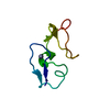
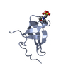
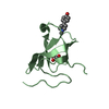
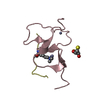
| Entry | Database: PDB / ID: 1pml | ||||||
|---|---|---|---|---|---|---|---|
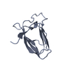
| Title | KRINGLE-KRINGLE INTERACTIONS IN MULTIMER KRINGLE STRUCTURES | ||||||
 Components Components | TISSUE PLASMINOGEN ACTIVATOR KRINGLE 2 | ||||||
 Keywords Keywords | HYDROLASE(SERINE PROTEASE) | ||||||
| Function / homology |  Function and homology information Function and homology informationt-plasminogen activator / prevention of polyspermy / trans-synaptic signaling by BDNF, modulating synaptic transmission / Signaling by PDGF / negative regulation of plasminogen activation / Dissolution of Fibrin Clot / smooth muscle cell migration / plasminogen activation / platelet-derived growth factor receptor signaling pathway / negative regulation of fibrinolysis ...t-plasminogen activator / prevention of polyspermy / trans-synaptic signaling by BDNF, modulating synaptic transmission / Signaling by PDGF / negative regulation of plasminogen activation / Dissolution of Fibrin Clot / smooth muscle cell migration / plasminogen activation / platelet-derived growth factor receptor signaling pathway / negative regulation of fibrinolysis / serine protease inhibitor complex / fibrinolysis / negative regulation of proteolysis / secretory granule / protein modification process / phosphoprotein binding / Schaffer collateral - CA1 synapse / apical part of cell / blood coagulation / response to hypoxia / signaling receptor binding / serine-type endopeptidase activity / glutamatergic synapse / cell surface / proteolysis / : / extracellular exosome / extracellular region / cytoplasm Similarity search - Function | ||||||
| Biological species |  Homo sapiens (human) Homo sapiens (human) | ||||||
| Method |  X-RAY DIFFRACTION / Resolution: 2.38 Å X-RAY DIFFRACTION / Resolution: 2.38 Å | ||||||
 Authors Authors | Padmanabhan, K. / Tulinsky, A. | ||||||
 Citation Citation | Journal: Protein Sci. / Year: 1994 Title: Kringle-kringle interactions in multimer kringle structures. Authors: Padmanabhan, K. / Wu, T.P. / Ravichandran, K.G. / Tulinsky, A. #1: Journal: Biochemistry / Year: 1992Title: Crystal Structure of the Kringle 2 Domain of Tissue Plasminogen Activator at 2.4 A Resolution Authors: De Vos, A.M. / Ultsch, M.H. / Kelly, R.F. / Padmanabhan, K. / Tulinsky, A. / Westbrook, M.L. / Kossiakoff, A.A. #2: Journal: Biochemistry / Year: 1991Title: Crystal and Molecular Structure of Human Plasminogen Kringle 4 Refined to 1.9A Resolution Authors: Mulichak, A.M. / Tulinsky, A. / Ravichandran, K.G. #3: Journal: Biochemistry / Year: 1991Title: The Refined Structure of the Epsilon-Aminocaproic Acid Complex of Human Plasminogen Kringle 4 Authors: Wu, T.-P. / Padmanabhan, K. / Tulinsky, A. / Mulichak, A.M. | ||||||
| History |
|
- Structure visualization
| Structure viewer | Molecule: MolmilJmol/JSmol |
|---|
- Downloads & links
-Download
| PDBx/mmCIF format | 1pml.cif.gz | 64.2 KB | Display | PDBx/mmCIF format |
|---|---|---|---|---|
| PDB format | pdb1pml.ent.gz | 46.9 KB | Display | PDB format |
| PDBx/mmJSON format | 1pml.json.gz | Tree view | PDBx/mmJSON format | |
| Others |  Other downloads Other downloads |
-Validation report
| Arichive directory | https://data.pdbj.org/pub/pdb/validation_reports/pm/1pmlftp://data.pdbj.org/pub/pdb/validation_reports/pm/1pml | HTTPS FTP |
|---|
-Related structure data


-Links
 PDBj
PDBj


- Assembly
| Deposited unit | 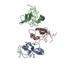
| ||||||||
|---|---|---|---|---|---|---|---|---|---|
| 1 | 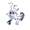
| ||||||||
| 2 | 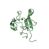
| ||||||||
| 3 | 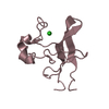
| ||||||||
| Unit cell |
| ||||||||
| Noncrystallographic symmetry (NCS) | NCS oper: (Code: given Matrix: (0.99997, -0.0059, 0.024), Vector: Details | THE TRANSFORMATION PRESENTED ON *MTRIX* RECORDS BELOW WILL YIELD APPROXIMATE COORDINATES FOR CHAIN A WHEN APPLIED TO CHAIN B. | |
-Components
| #1: Protein | Mass: 9482.614 Da / Num. of mol.: 3 Source method: isolated from a genetically manipulated source Source: (gene. exp.) Homo sapiens (human) / References: UniProt: P00750, t-plasminogen activator#2: Chemical |   Mass: 35.453 Da / Num. of mol.: 3 / Source method: obtained synthetically / Formula: Cl Mass: 35.453 Da / Num. of mol.: 3 / Source method: obtained synthetically / Formula: Cl#3: Water | ChemComp-HOH / |  Mass: 18.015 Da / Num. of mol.: 190 / Source method: isolated from a natural source / Formula: H2O Mass: 18.015 Da / Num. of mol.: 190 / Source method: isolated from a natural source / Formula: H2OHas protein modification | Y | |
|---|
-Experimental details
-Experiment
| Experiment | Method: X-RAY DIFFRACTION |
|---|
- Sample preparation
| Crystal | Density Matthews: 2.73 Å3/Da / Density % sol: 54.96 % | ||||||||||||||||||||
|---|---|---|---|---|---|---|---|---|---|---|---|---|---|---|---|---|---|---|---|---|---|
| Crystal grow | *PLUS pH: 8 / Method: vapor diffusion | ||||||||||||||||||||
| Components of the solutions | *PLUS
|
-Data collection
| Radiation | Scattering type: x-ray |
|---|---|
| Radiation wavelength | Relative weight: 1 |
| Reflection | *PLUS Highest resolution: 2.38 Å / Num. obs: 11621 / % possible obs: 91 % / Num. measured all: 30076 / Rmerge(I) obs: 0.07 |
| Reflection shell | *PLUS Highest resolution: 2.38 Å / Lowest resolution: 2.5 Å / % possible obs: 52 % / Num. possible: 2105 / Num. unique obs: 1101 / Mean I/σ(I) obs: 3.7 |
- Processing
| Software | Name: PROFFT / Classification: refinement | |||||||||||||||||||||||||||||||||||||||||||||||||||||||||||||||
|---|---|---|---|---|---|---|---|---|---|---|---|---|---|---|---|---|---|---|---|---|---|---|---|---|---|---|---|---|---|---|---|---|---|---|---|---|---|---|---|---|---|---|---|---|---|---|---|---|---|---|---|---|---|---|---|---|---|---|---|---|---|---|---|---|
| Refinement | Resolution: 2.38→8 Å / σ(F): 4 Details: NO ELECTRON DENSITY WAS OBSERVED FOR THE INTERKRINGLE RESIDUES -3 - -2 IN MOLECULE A AND MOLECULE B AND -3 - -2 AND 82 IN MOLECULE C.
| |||||||||||||||||||||||||||||||||||||||||||||||||||||||||||||||
| Refinement step | Cycle: LAST / Resolution: 2.38→8 Å
| |||||||||||||||||||||||||||||||||||||||||||||||||||||||||||||||
| Refine LS restraints |
| |||||||||||||||||||||||||||||||||||||||||||||||||||||||||||||||
| Software | *PLUS Name: PROFFT / Classification: refinement | |||||||||||||||||||||||||||||||||||||||||||||||||||||||||||||||
| Refinement | *PLUS Rfactor obs: 0.145 | |||||||||||||||||||||||||||||||||||||||||||||||||||||||||||||||
| Solvent computation | *PLUS | |||||||||||||||||||||||||||||||||||||||||||||||||||||||||||||||
| Displacement parameters | *PLUS Biso mean: 21.7 Å2 | |||||||||||||||||||||||||||||||||||||||||||||||||||||||||||||||
| Refine LS restraints | *PLUS
|
