 Movie
Movie Controller

[English] 日本語
 Yorodumi
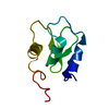
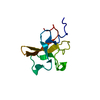
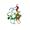
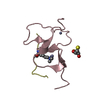
- PDB-7a9a: Crystal structure of rubredoxin B (Rv3250c) from Mycobacterium tu... -
+ Open data

- Basic information
| Entry | Database: PDB / ID: 7a9a | |||||||||
|---|---|---|---|---|---|---|---|---|---|---|
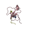
| Title | Crystal structure of rubredoxin B (Rv3250c) from Mycobacterium tuberculosis | |||||||||
 Components Components | Rubredoxin | |||||||||
 Keywords Keywords | ELECTRON TRANSPORT / IRON SULFUR / METALLOPROTEIN / CYTOCHROME P450 REDOX PARTNER / CYP / CYP124 / CYP125 / CYP142 | |||||||||
| Function / homology |  Function and homology information Function and homology informationalkane catabolic process / electron transfer activity / iron ion binding Similarity search - Function | |||||||||
| Biological species |   Mycobacterium tuberculosis (bacteria) Mycobacterium tuberculosis (bacteria) | |||||||||
| Method |  X-RAY DIFFRACTION / SYNCHROTRON / MOLECULAR REPLACEMENT / Resolution: 1.17 Å X-RAY DIFFRACTION / SYNCHROTRON / MOLECULAR REPLACEMENT / Resolution: 1.17 Å | |||||||||
 Authors Authors | Vakhrameev, D. / Kavaleuski, A. / Bukhdruker, S. / Marin, E. / Sushko, T. / Grabovec, I.P. / Gilep, A. / Strushkevich, N. / Borshchevskiy, V. | |||||||||
| Funding support |  Russian Federation, Belarus, 2items Russian Federation, Belarus, 2items
| |||||||||
 Citation Citation | Journal: Bioorg.Chem. / Year: 2021 Title: A new twist of rubredoxin function in M. tuberculosis. Authors: Sushko, T. / Kavaleuski, A. / Grabovec, I. / Kavaleuskaya, A. / Vakhrameev, D. / Bukhdruker, S. / Marin, E. / Kuzikov, A. / Masamrekh, R. / Shumyantseva, V. / Tsumoto, K. / Borshchevskiy, V. ...Authors: Sushko, T. / Kavaleuski, A. / Grabovec, I. / Kavaleuskaya, A. / Vakhrameev, D. / Bukhdruker, S. / Marin, E. / Kuzikov, A. / Masamrekh, R. / Shumyantseva, V. / Tsumoto, K. / Borshchevskiy, V. / Gilep, A. / Strushkevich, N. | |||||||||
| History |
|
- Structure visualization
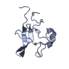
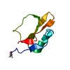
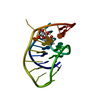
| Structure viewer | Molecule: MolmilJmol/JSmol |
|---|
- Downloads & links
-Download
| PDBx/mmCIF format | 7a9a.cif.gz | 334.5 KB | Display | PDBx/mmCIF format |
|---|---|---|---|---|
| PDB format | pdb7a9a.ent.gz | 267.6 KB | Display | PDB format |
| PDBx/mmJSON format | 7a9a.json.gz | Tree view | PDBx/mmJSON format | |
| Others |  Other downloads Other downloads |
-Validation report
| Arichive directory | https://data.pdbj.org/pub/pdb/validation_reports/a9/7a9aftp://data.pdbj.org/pub/pdb/validation_reports/a9/7a9a | HTTPS FTP |
|---|
-Related structure data
| Related structure data | 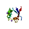 2pyaS S: Starting model for refinement |
|---|---|
| Similar structure data |

-Links
 PDBj
PDBj
- Assembly
| Deposited unit | 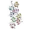
| ||||||||||||
|---|---|---|---|---|---|---|---|---|---|---|---|---|---|
| 1 | 
| ||||||||||||
| 2 | 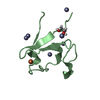
| ||||||||||||
| 3 | 
| ||||||||||||
| 4 | 
| ||||||||||||
| 5 | 
| ||||||||||||
| 6 | 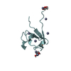
| ||||||||||||
| 7 | 
| ||||||||||||
| 8 | 
| ||||||||||||
| Unit cell |
|
-Components
-Protein , 1 types, 8 molecules ABCDEFGH
| #1: Protein | Mass: 6802.464 Da / Num. of mol.: 8 Source method: isolated from a genetically manipulated source Source: (gene. exp.) Mycobacterium tuberculosis (strain ATCC 25618 / H37Rv) (bacteria)Strain: ATCC 25618 / H37Rv / Gene: rubB, Rv3250c / Plasmid: pET11a / Production host: |
|---|
-Non-polymers , 7 types, 639 molecules 


| #2: Chemical | ChemComp-FE /  Mass: 55.845 Da / Num. of mol.: 8 / Source method: isolated from a natural source / Formula: Fe Mass: 55.845 Da / Num. of mol.: 8 / Source method: isolated from a natural source / Formula: Fe#3: Chemical | ChemComp-ZN /  Mass: 65.409 Da / Num. of mol.: 40 / Source method: obtained synthetically / Formula: Zn Mass: 65.409 Da / Num. of mol.: 40 / Source method: obtained synthetically / Formula: Zn#4: Chemical |  Mass: 62.068 Da / Num. of mol.: 3 / Source method: obtained synthetically / Formula: C2H6O2 Mass: 62.068 Da / Num. of mol.: 3 / Source method: obtained synthetically / Formula: C2H6O2#5: Chemical | ChemComp-GOL /  Mass: 92.094 Da / Num. of mol.: 4 / Source method: obtained synthetically / Formula: C3H8O3 Mass: 92.094 Da / Num. of mol.: 4 / Source method: obtained synthetically / Formula: C3H8O3#6: Chemical | ChemComp-CL / |  Mass: 35.453 Da / Num. of mol.: 1 / Source method: obtained synthetically / Formula: Cl Mass: 35.453 Da / Num. of mol.: 1 / Source method: obtained synthetically / Formula: Cl#7: Chemical | ChemComp-PEG /  Mass: 106.120 Da / Num. of mol.: 4 / Source method: obtained synthetically / Formula: C4H10O3 Mass: 106.120 Da / Num. of mol.: 4 / Source method: obtained synthetically / Formula: C4H10O3#8: Water | ChemComp-HOH / | Mass: 18.015 Da / Num. of mol.: 579 / Source method: isolated from a natural source / Formula: H2O |
|---|
-Details
| Has ligand of interest | N |
|---|
-Experimental details
-Experiment
| Experiment | Method: X-RAY DIFFRACTION / Number of used crystals: 1 |
|---|
- Sample preparation
| Crystal | Density Matthews: 1.82 Å3/Da / Density % sol: 32.38 % |
|---|---|
| Crystal grow | Temperature: 293 K / Method: vapor diffusion, sitting drop / pH: 7.5 / Details: 100 mM Tris-HCl, 50 mM zinc acetate, 25 % PEG 3350 |
-Data collection
| Diffraction | Mean temperature: 100 K / Serial crystal experiment: N |
|---|---|
| Diffraction source | Source: SYNCHROTRON / Site: ESRF  / Beamline: ID30B / Wavelength: 0.97625 Å / Beamline: ID30B / Wavelength: 0.97625 Å |
| Detector | Type: DECTRIS PILATUS3 6M / Detector: PIXEL / Date: Jul 8, 2018 |
| Radiation | Protocol: SINGLE WAVELENGTH / Monochromatic (M) / Laue (L): M / Scattering type: x-ray |
| Radiation wavelength | Wavelength: 0.97625 Å / Relative weight: 1 |
| Reflection | Resolution: 1.17→47.95 Å / Num. obs: 119411 / % possible obs: 93.18 % / Redundancy: 3.5 % / Biso Wilson estimate: 9.13 Å2 / CC1/2: 0.994 / Rmerge(I) obs: 0.135 / Rpim(I) all: 0.086 / Rrim(I) all: 0.16 / Net I/σ(I): 4.69 |
| Reflection shell | Resolution: 1.17→1.31 Å / Redundancy: 3.5 % / Rmerge(I) obs: 2.077 / Mean I/σ(I) obs: 0.55 / Num. unique obs: 119411 / CC1/2: 0.295 / Rpim(I) all: 1.312 / Rrim(I) all: 2.466 / % possible all: 90.95 |
- Processing
| Software |
| ||||||||||||||||||||||||
|---|---|---|---|---|---|---|---|---|---|---|---|---|---|---|---|---|---|---|---|---|---|---|---|---|---|
| Refinement | Method to determine structure: MOLECULAR REPLACEMENT Starting model: 2PYA Resolution: 1.17→47.95 Å / SU ML: 0.1186 / Cross valid method: FREE R-VALUE / σ(F): 1.94 / Phase error: 23.9895 Stereochemistry target values: GeoStd + Monomer Library + CDL v1.2
| ||||||||||||||||||||||||
| Solvent computation | Shrinkage radii: 0.9 Å / VDW probe radii: 1.11 Å / Solvent model: FLAT BULK SOLVENT MODEL | ||||||||||||||||||||||||
| Displacement parameters | Biso mean: 13.47 Å2 | ||||||||||||||||||||||||
| Refinement step | Cycle: LAST / Resolution: 1.17→47.95 Å
| ||||||||||||||||||||||||
| Refine LS restraints |
| ||||||||||||||||||||||||
| LS refinement shell | Resolution: 1.17→47.95 Å |
