 Movie
Movie Controller

[English] 日本語
 Yorodumi
- PDB-6q72: Crystal structure of the alanine racemase from Bacillus subtilis ... -
+ Open data

- Basic information
| Entry | Database: PDB / ID: 6q72 | ||||||
|---|---|---|---|---|---|---|---|
| Title | Crystal structure of the alanine racemase from Bacillus subtilis in the presence of only PEG 4000 and Magnesium chloride in the crystallization condition | ||||||
 Components Components | Alanine racemase 2 | ||||||
 Keywords Keywords | ISOMERASE / racemase / alanine racemase | ||||||
| Function / homology |  Function and homology information Function and homology informationalanine racemase / D-alanine biosynthetic process / alanine racemase activity / peptidoglycan biosynthetic process / pyridoxal phosphate binding / cytosol Similarity search - Function | ||||||
| Biological species |  | ||||||
| Method |  X-RAY DIFFRACTION / SYNCHROTRON / MOLECULAR REPLACEMENT / Resolution: 3 Å X-RAY DIFFRACTION / SYNCHROTRON / MOLECULAR REPLACEMENT / Resolution: 3 Å | ||||||
 Authors Authors | Bernardo-Garcia, N. / Gago, F. / Hermoso, J.A. | ||||||
 Citation Citation | Journal: Org.Biomol.Chem. / Year: 2019 Title: Cold-induced aldimine bond cleavage by Tris in Bacillus subtilis alanine racemase. Authors: Bernardo-Garcia, N. / Sanchez-Murcia, P.A. / Espaillat, A. / Martinez-Caballero, S. / Cava, F. / Hermoso, J.A. / Gago, F. | ||||||
| History |
|
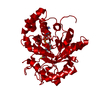
- Structure visualization
| Structure viewer | Molecule: MolmilJmol/JSmol |
|---|
- Downloads & links
-Download
| PDBx/mmCIF format | 6q72.cif.gz | 305.7 KB | Display | PDBx/mmCIF format |
|---|---|---|---|---|
| PDB format | pdb6q72.ent.gz | 249.9 KB | Display | PDB format |
| PDBx/mmJSON format | 6q72.json.gz | Tree view | PDBx/mmJSON format | |
| Others |  Other downloads Other downloads |
-Validation report
| Arichive directory | https://data.pdbj.org/pub/pdb/validation_reports/q7/6q72ftp://data.pdbj.org/pub/pdb/validation_reports/q7/6q72 | HTTPS FTP |
|---|
-Related structure data
| Related structure data | 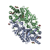 5irpC  6q70C 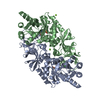 6q71C  5rip C: citing same article ( S: Starting model for refinement |
|---|---|
| Similar structure data |


-Links
 PDBj
PDBj- Assembly
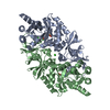
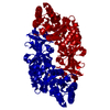
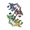
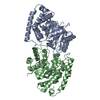
| Deposited unit | 
| ||||||||
|---|---|---|---|---|---|---|---|---|---|
| 1 | 
| ||||||||
| 2 | 
| ||||||||
| Unit cell |
|
-Components
| #1: Protein | Mass: 43708.840 Da / Num. of mol.: 4 Source method: isolated from a genetically manipulated source Source: (gene. exp.) #2: Chemical | ChemComp-PLP /   Mass: 247.142 Da / Num. of mol.: 4 / Source method: obtained synthetically / Formula: C8H10NO6P Mass: 247.142 Da / Num. of mol.: 4 / Source method: obtained synthetically / Formula: C8H10NO6P#3: Chemical | ChemComp-CL /   Mass: 35.453 Da / Num. of mol.: 4 Mass: 35.453 Da / Num. of mol.: 4Source method: isolated from a genetically manipulated source Formula: Cl #4: Water | ChemComp-HOH / |  Mass: 18.015 Da / Num. of mol.: 7 / Source method: isolated from a natural source / Formula: H2O Mass: 18.015 Da / Num. of mol.: 7 / Source method: isolated from a natural source / Formula: H2O |
|---|
-Experimental details
-Experiment
| Experiment | Method: X-RAY DIFFRACTION / Number of used crystals: 1 |
|---|
- Sample preparation
| Crystal | Density Matthews: 2.23 Å3/Da / Density % sol: 44.74 % |
|---|---|
| Crystal grow | Temperature: 290 K / Method: microbatch / Details: 15% PEG 4000, 0.2 MgCl2 |
-Data collection
| Diffraction | Mean temperature: 100 K / Serial crystal experiment: N |
|---|---|
| Diffraction source | Source: SYNCHROTRON / Site: ALBA  / Beamline: XALOC / Wavelength: 0.97926 Å / Beamline: XALOC / Wavelength: 0.97926 Å |
| Detector | Type: DECTRIS PILATUS 6M / Detector: PIXEL / Date: Jun 23, 2016 |
| Radiation | Protocol: SINGLE WAVELENGTH / Monochromatic (M) / Laue (L): M / Scattering type: x-ray |
| Radiation wavelength | Wavelength: 0.97926 Å / Relative weight: 1 |
| Reflection | Resolution: 2.85→46.61 Å / Num. obs: 35532 / % possible obs: 99.5 % / Redundancy: 3.2 % / Rmerge(I) obs: 0.048 / Net I/σ(I): 5.7 |
| Reflection shell | Resolution: 2.85→2.99 Å |
- Processing
| Software |
| ||||||||||||||||||||||||||||||||||||||||||||||||||||||||||||||||||||||||||||||||||||
|---|---|---|---|---|---|---|---|---|---|---|---|---|---|---|---|---|---|---|---|---|---|---|---|---|---|---|---|---|---|---|---|---|---|---|---|---|---|---|---|---|---|---|---|---|---|---|---|---|---|---|---|---|---|---|---|---|---|---|---|---|---|---|---|---|---|---|---|---|---|---|---|---|---|---|---|---|---|---|---|---|---|---|---|---|---|
| Refinement | Method to determine structure: MOLECULAR REPLACEMENT Starting model: 5RIP 5rip Resolution: 3→45.32 Å / SU ML: 0.43 / Cross valid method: FREE R-VALUE / σ(F): 1.33 / Phase error: 31.59
| ||||||||||||||||||||||||||||||||||||||||||||||||||||||||||||||||||||||||||||||||||||
| Solvent computation | Shrinkage radii: 0.9 Å / VDW probe radii: 1.11 Å | ||||||||||||||||||||||||||||||||||||||||||||||||||||||||||||||||||||||||||||||||||||
| Refinement step | Cycle: LAST / Resolution: 3→45.32 Å
| ||||||||||||||||||||||||||||||||||||||||||||||||||||||||||||||||||||||||||||||||||||
| Refine LS restraints |
| ||||||||||||||||||||||||||||||||||||||||||||||||||||||||||||||||||||||||||||||||||||
| LS refinement shell |
|
