 Movie
Movie Controller

[English] 日本語
 Yorodumi
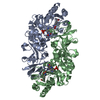
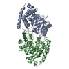
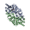
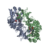
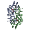
- PDB-1vfh: Crystal structure of alanine racemase from D-cycloserine producin... -
+ Open data

- Basic information
| Entry | Database: PDB / ID: 1vfh | ||||||
|---|---|---|---|---|---|---|---|
| Title | Crystal structure of alanine racemase from D-cycloserine producing Streptomyces lavendulae | ||||||
 Components Components | alanine racemase | ||||||
 Keywords Keywords | ISOMERASE / TIM-barrel / Greek-key motief | ||||||
| Function / homology |  Function and homology information Function and homology informationalanine racemase / D-alanine biosynthetic process / alanine racemase activity / peptidoglycan biosynthetic process / pyridoxal phosphate binding / cytosol Similarity search - Function | ||||||
| Biological species |  Streptomyces lavendulae (bacteria) Streptomyces lavendulae (bacteria) | ||||||
| Method |  X-RAY DIFFRACTION / MOLECULAR REPLACEMENT / Resolution: 2 Å X-RAY DIFFRACTION / MOLECULAR REPLACEMENT / Resolution: 2 Å | ||||||
 Authors Authors | Noda, M. / Matoba, Y. / Kumagai, T. / Sugiyama, M. | ||||||
 Citation Citation | Journal: J.Biol.Chem. / Year: 2004 Title: Structural evidence that alanine racemase from a D-cycloserine-producing microorganism exhibits resistance to its own product. Authors: Noda, M. / Matoba, Y. / Kumagai, T. / Sugiyama, M. #1: Journal: J.Biol.Chem. / Year: 2004 Title: Self-protection mechanism in D-cycloserine-producing streptomyces lavendulae: Gene cloning, characterization, and kinetics of its alanine racemase and D-alanyl-D-alanine ligase. which are ...Title: Self-protection mechanism in D-cycloserine-producing streptomyces lavendulae: Gene cloning, characterization, and kinetics of its alanine racemase and D-alanyl-D-alanine ligase. which are traget enyzmes of D-cycloserine Authors: Noda, M. / Kawahara, Y. / Ichikawa, A. / Matoba, Y. / Matsuo, H. / Lee, D.G. / Kumagai, T. / Sugiyama, M. | ||||||
| History |
|
- Structure visualization
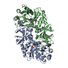
| Structure viewer | Molecule: MolmilJmol/JSmol |
|---|
- Downloads & links
-Download
| PDBx/mmCIF format | 1vfh.cif.gz | 86.7 KB | Display | PDBx/mmCIF format |
|---|---|---|---|---|
| PDB format | pdb1vfh.ent.gz | 63.9 KB | Display | PDB format |
| PDBx/mmJSON format | 1vfh.json.gz | Tree view | PDBx/mmJSON format | |
| Others |  Other downloads Other downloads |
-Validation report
| Arichive directory | https://data.pdbj.org/pub/pdb/validation_reports/vf/1vfhftp://data.pdbj.org/pub/pdb/validation_reports/vf/1vfh | HTTPS FTP |
|---|
-Related structure data
| Related structure data |  1vfsC  1vftC  1sftS C: citing same article ( S: Starting model for refinement |
|---|---|
| Similar structure data |


-Links
 PDBj
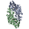
PDBj- Assembly
| Deposited unit | 
| ||||||||
|---|---|---|---|---|---|---|---|---|---|
| 1 | 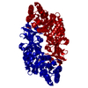
| ||||||||
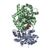
| Unit cell |
| ||||||||
| Details | The second part of the biological assembly is generated by the two fold axis: -x, y, 1-z. |
-Components
| #1: Protein | Mass: 41049.160 Da / Num. of mol.: 1 Source method: isolated from a genetically manipulated source Source: (gene. exp.) Streptomyces lavendulae (bacteria) / Gene: alr / Plasmid: pET-alr / Production host: |
|---|---|
| #2: Chemical | ChemComp-PLP /   Mass: 247.142 Da / Num. of mol.: 1 / Source method: obtained synthetically / Formula: C8H10NO6P Mass: 247.142 Da / Num. of mol.: 1 / Source method: obtained synthetically / Formula: C8H10NO6P |
| #3: Water | ChemComp-HOH /  Mass: 18.015 Da / Num. of mol.: 74 / Source method: isolated from a natural source / Formula: H2O Mass: 18.015 Da / Num. of mol.: 74 / Source method: isolated from a natural source / Formula: H2O |
-Experimental details
-Experiment
| Experiment | Method: X-RAY DIFFRACTION / Number of used crystals: 1 |
|---|
- Sample preparation
| Crystal | Density Matthews: 2.41 Å3/Da / Density % sol: 48.92 % |
|---|---|
| Crystal grow | Temperature: 298 K / Method: vapor diffusion, sitting drop / pH: 8.5 Details: Ammonium sulfate, pH 8.5, VAPOR DIFFUSION, SITTING DROP, temperature 298K |
-Data collection
| Diffraction | Mean temperature: 298 K |
|---|---|
| Diffraction source | Source: ROTATING ANODE / Type: RIGAKU / Wavelength: 1.5418 Å |
| Detector | Type: RIGAKU RAXIS / Detector: IMAGE PLATE / Date: May 22, 2003 / Details: mirrors |
| Radiation | Monochromator: mirror / Protocol: SINGLE WAVELENGTH / Monochromatic (M) / Laue (L): M / Scattering type: x-ray |
| Radiation wavelength | Wavelength: 1.5418 Å / Relative weight: 1 |
| Reflection | Resolution: 2→100 Å / Num. all: 24889 / Num. obs: 24889 / % possible obs: 94 % / Observed criterion σ(F): 0 / Observed criterion σ(I): 0 / Redundancy: 2.9 % / Biso Wilson estimate: 24.1 Å2 / Rmerge(I) obs: 0.111 / Rsym value: 0.092 / Net I/σ(I): 6.7 |
| Reflection shell | Resolution: 2→2.11 Å / Redundancy: 2.1 % / Rmerge(I) obs: 0.609 / Mean I/σ(I) obs: 1.5 / Num. unique all: 6953 / Rsym value: 0.475 / % possible all: 86.9 |
- Processing
| Software |
| ||||||||||||||||||||||||||||||||||||
|---|---|---|---|---|---|---|---|---|---|---|---|---|---|---|---|---|---|---|---|---|---|---|---|---|---|---|---|---|---|---|---|---|---|---|---|---|---|
| Refinement | Method to determine structure: MOLECULAR REPLACEMENT Starting model: PDB entry 1SFT Resolution: 2→30 Å / Rfactor Rfree error: 0.007 / Data cutoff high absF: 10000000 / Data cutoff low absF: 0.001 / Isotropic thermal model: RESTRAINED / Cross valid method: THROUGHOUT / σ(F): 2 / σ(I): 1 / Stereochemistry target values: Engh & Huber
| ||||||||||||||||||||||||||||||||||||
| Displacement parameters | Biso mean: 33 Å2
| ||||||||||||||||||||||||||||||||||||
| Refine analyze |
| ||||||||||||||||||||||||||||||||||||
| Refinement step | Cycle: LAST / Resolution: 2→30 Å
| ||||||||||||||||||||||||||||||||||||
| Refine LS restraints |
| ||||||||||||||||||||||||||||||||||||
| LS refinement shell | Resolution: 2→2.13 Å / Rfactor Rfree error: 0.029 / Total num. of bins used: 6
| ||||||||||||||||||||||||||||||||||||
| Xplor file |
|
