 Movie
Movie Controller

[English] 日本語
 Yorodumi
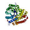
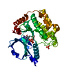
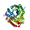
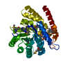
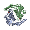
- PDB-5czj: Crystal structure of HypD, a 1-pyrroline-4-hydroxy-2-carboxylate ... -
+ Open data

- Basic information
| Entry | Database: PDB / ID: 5czj | ||||||
|---|---|---|---|---|---|---|---|
| Title | Crystal structure of HypD, a 1-pyrroline-4-hydroxy-2-carboxylate deaminase from Sinorhizobium meliloti | ||||||
 Components Components | Dihydrodipicolinate synthase | ||||||
 Keywords Keywords | HYDROLASE / N-acetylneuraminate lyase sub-family / (alpha/beta)8 barrel / TIM barrel / 4-hydroxy-proline metabolism | ||||||
| Function / homology |  Function and homology information Function and homology informationdihydrodipicolinate synthase / 4-hydroxy-tetrahydrodipicolinate synthase activity Similarity search - Function | ||||||
| Biological species |  Rhizobium meliloti (bacteria) Rhizobium meliloti (bacteria) | ||||||
| Method |  X-RAY DIFFRACTION / MOLECULAR REPLACEMENT / Resolution: 1.92 Å X-RAY DIFFRACTION / MOLECULAR REPLACEMENT / Resolution: 1.92 Å | ||||||
 Authors Authors | Stogios, P.J. / Xu, X. / Savchenko, A. | ||||||
 Citation Citation | Journal: J.Bacteriol. / Year: 2016 Title: l-Hydroxyproline and d-Proline Catabolism in Sinorhizobium meliloti. Authors: Chen, S. / White, C.E. / diCenzo, G.C. / Zhang, Y. / Stogios, P.J. / Savchenko, A. / Finan, T.M. | ||||||
| History |
|
- Structure visualization
| Structure viewer | Molecule: MolmilJmol/JSmol |
|---|
- Downloads & links
-Download
| PDBx/mmCIF format | 5czj.cif.gz | 260.8 KB | Display | PDBx/mmCIF format |
|---|---|---|---|---|
| PDB format | pdb5czj.ent.gz | 212.7 KB | Display | PDB format |
| PDBx/mmJSON format | 5czj.json.gz | Tree view | PDBx/mmJSON format | |
| Others |  Other downloads Other downloads |
-Validation report
| Arichive directory | https://data.pdbj.org/pub/pdb/validation_reports/cz/5czjftp://data.pdbj.org/pub/pdb/validation_reports/cz/5czj | HTTPS FTP |
|---|
-Related structure data
| Related structure data |  2hmcS S: Starting model for refinement |
|---|---|
| Similar structure data |


-Links
 PDBj
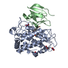
PDBj- Assembly
| Deposited unit | 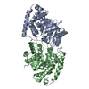
| ||||||||
|---|---|---|---|---|---|---|---|---|---|
| 1 | 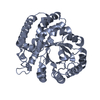
| ||||||||
| 2 | 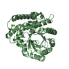
| ||||||||
| 3 |
| ||||||||
| 4 | 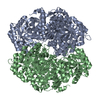
| ||||||||
| Unit cell |
|
-Components
| #1: Protein | Mass: 34485.527 Da / Num. of mol.: 2 Source method: isolated from a genetically manipulated source Source: (gene. exp.) Rhizobium meliloti (strain 1021) (bacteria)Strain: 1021 / Gene: hypD, SM_b20259 / Plasmid: p15TV-LIC / Production host: #2: Water | ChemComp-HOH / |  Mass: 18.015 Da / Num. of mol.: 539 / Source method: isolated from a natural source / Formula: H2O Mass: 18.015 Da / Num. of mol.: 539 / Source method: isolated from a natural source / Formula: H2O |
|---|
-Experimental details
-Experiment
| Experiment | Method: X-RAY DIFFRACTION / Number of used crystals: 1 |
|---|
- Sample preparation
| Crystal | Density Matthews: 2.35 Å3/Da / Density % sol: 47.7 % |
|---|---|
| Crystal grow | Temperature: 298 K / Method: vapor diffusion, hanging drop / pH: 7 Details: 0.5 microL of 19.5 mg/mL protein solution (including 20% (w/v) glycerol) mixed with 0.5 microL of reservoir solution (0.2 M potassium/sodium tartrate, 20% (w/v) PEG3350). |
-Data collection
| Diffraction | Mean temperature: 100 K |
|---|---|
| Diffraction source | Source: ROTATING ANODE / Type: RIGAKU FR-E SUPERBRIGHT / Wavelength: 1.5418 Å |
| Detector | Type: RIGAKU RAXIS HTC / Detector: IMAGE PLATE / Date: Oct 16, 2009 |
| Radiation | Protocol: SINGLE WAVELENGTH / Monochromatic (M) / Laue (L): M / Scattering type: x-ray |
| Radiation wavelength | Wavelength: 1.5418 Å / Relative weight: 1 |
| Reflection | Resolution: 1.92→29.431 Å / Num. obs: 49734 / % possible obs: 100 % / Redundancy: 21.3 % / Rmerge(I) obs: 0.071 / Net I/σ(I): 36.88 |
| Reflection shell | Resolution: 1.92→1.95 Å / Redundancy: 19.8 % / Rmerge(I) obs: 0.583 / % possible all: 100 |
- Processing
| Software |
| |||||||||||||||||||||||||||||||||||||||||||||||||||||||||||||||||||||||||||||||||||||||||||||||||||||||||||||||||||||||||||||||||||||||||||||||||||||||||||||||||||||||||||||||||||||||||||||||||||||||||||||||||||||||||||||||||||||||||||||||||||||||||||||||||||||||||||||||||||
|---|---|---|---|---|---|---|---|---|---|---|---|---|---|---|---|---|---|---|---|---|---|---|---|---|---|---|---|---|---|---|---|---|---|---|---|---|---|---|---|---|---|---|---|---|---|---|---|---|---|---|---|---|---|---|---|---|---|---|---|---|---|---|---|---|---|---|---|---|---|---|---|---|---|---|---|---|---|---|---|---|---|---|---|---|---|---|---|---|---|---|---|---|---|---|---|---|---|---|---|---|---|---|---|---|---|---|---|---|---|---|---|---|---|---|---|---|---|---|---|---|---|---|---|---|---|---|---|---|---|---|---|---|---|---|---|---|---|---|---|---|---|---|---|---|---|---|---|---|---|---|---|---|---|---|---|---|---|---|---|---|---|---|---|---|---|---|---|---|---|---|---|---|---|---|---|---|---|---|---|---|---|---|---|---|---|---|---|---|---|---|---|---|---|---|---|---|---|---|---|---|---|---|---|---|---|---|---|---|---|---|---|---|---|---|---|---|---|---|---|---|---|---|---|---|---|---|---|---|---|---|---|---|---|---|---|---|---|---|---|---|---|---|---|---|---|---|---|---|---|---|---|---|---|---|---|---|---|---|---|---|---|---|---|---|---|---|---|---|---|---|---|---|---|---|---|---|
| Refinement | Method to determine structure: MOLECULAR REPLACEMENT Starting model: 2hmc Resolution: 1.92→29.431 Å / SU ML: 0.34 / Cross valid method: FREE R-VALUE / σ(F): 1.38 / Phase error: 33.75 / Stereochemistry target values: ML
| |||||||||||||||||||||||||||||||||||||||||||||||||||||||||||||||||||||||||||||||||||||||||||||||||||||||||||||||||||||||||||||||||||||||||||||||||||||||||||||||||||||||||||||||||||||||||||||||||||||||||||||||||||||||||||||||||||||||||||||||||||||||||||||||||||||||||||||||||||
| Solvent computation | Shrinkage radii: 0.9 Å / VDW probe radii: 1.11 Å / Solvent model: FLAT BULK SOLVENT MODEL | |||||||||||||||||||||||||||||||||||||||||||||||||||||||||||||||||||||||||||||||||||||||||||||||||||||||||||||||||||||||||||||||||||||||||||||||||||||||||||||||||||||||||||||||||||||||||||||||||||||||||||||||||||||||||||||||||||||||||||||||||||||||||||||||||||||||||||||||||||
| Refinement step | Cycle: LAST / Resolution: 1.92→29.431 Å
| |||||||||||||||||||||||||||||||||||||||||||||||||||||||||||||||||||||||||||||||||||||||||||||||||||||||||||||||||||||||||||||||||||||||||||||||||||||||||||||||||||||||||||||||||||||||||||||||||||||||||||||||||||||||||||||||||||||||||||||||||||||||||||||||||||||||||||||||||||
| Refine LS restraints |
| |||||||||||||||||||||||||||||||||||||||||||||||||||||||||||||||||||||||||||||||||||||||||||||||||||||||||||||||||||||||||||||||||||||||||||||||||||||||||||||||||||||||||||||||||||||||||||||||||||||||||||||||||||||||||||||||||||||||||||||||||||||||||||||||||||||||||||||||||||
| LS refinement shell |
| |||||||||||||||||||||||||||||||||||||||||||||||||||||||||||||||||||||||||||||||||||||||||||||||||||||||||||||||||||||||||||||||||||||||||||||||||||||||||||||||||||||||||||||||||||||||||||||||||||||||||||||||||||||||||||||||||||||||||||||||||||||||||||||||||||||||||||||||||||
| Refinement TLS params. | Method: refined / Refine-ID: X-RAY DIFFRACTION
| |||||||||||||||||||||||||||||||||||||||||||||||||||||||||||||||||||||||||||||||||||||||||||||||||||||||||||||||||||||||||||||||||||||||||||||||||||||||||||||||||||||||||||||||||||||||||||||||||||||||||||||||||||||||||||||||||||||||||||||||||||||||||||||||||||||||||||||||||||
| Refinement TLS group |
|
