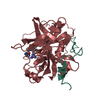
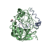
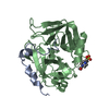
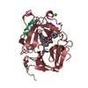
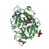
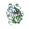
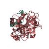
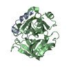
登録情報 データベース : PDB / ID : 6pxjタイトル Crystal structure of human thrombin mutant I16T Thrombin heavy chain Thrombin light chain キーワード / / 機能・相同性 分子機能 ドメイン・相同性 構成要素
/ / / / / / / / / / / / / / / / / / / / / / / / / / / / / / / / / / / / / / / / / / / / / / / / / / / / / / / / / / / / / / / / / / / / / / / / / / / / / / / / / / / / / / / / / / / / / / / / / / / / / / / / / / / / / / / / / / 生物種 Homo sapiens (ヒト)手法 / / 解像度 : 1.7 Å データ登録者 Stojanovski, B. / Chen, Z. / Koester, S.K. / Pelc, L.A. / Di Cera, E. 資金援助 組織 認可番号 国 National Institutes of Health/National Human Genome Research Institute (NIH/NHGRI)
履歴 登録 2019年7月26日 登録サイト / 処理サイト 改定 1.0 2019年12月18日 Provider / タイプ 改定 1.1 2023年10月11日 Group Data collection / Database references ... Data collection / Database references / Derived calculations / Refinement description カテゴリ chem_comp_atom / chem_comp_bond ... chem_comp_atom / chem_comp_bond / database_2 / pdbx_initial_refinement_model / pdbx_struct_conn_angle / struct_conn Item _database_2.pdbx_DOI / _database_2.pdbx_database_accession ... _database_2.pdbx_DOI / _database_2.pdbx_database_accession / _pdbx_struct_conn_angle.ptnr1_auth_seq_id / _pdbx_struct_conn_angle.ptnr3_auth_seq_id / _pdbx_struct_conn_angle.value / _struct_conn.pdbx_dist_value / _struct_conn.ptnr2_auth_seq_id 改定 1.2 2024年11月20日 Group カテゴリ / pdbx_modification_featureItem
すべて表示 表示を減らす
 ムービー
ムービー コントローラー


 データを開く
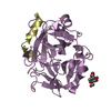
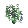
 基本情報
基本情報 要素
要素 キーワード
キーワード 機能・相同性情報
 Homo sapiens (ヒト)
 X線回折 /
  データ登録者
 米国, 1件
  引用
引用 構造の表示
構造の表示 ダウンロードとリンク
 その他のダウンロード


 PDBj
PDBj


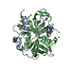
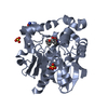
 集合体
集合体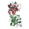

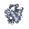
 Cricetulus griseus (モンゴルキヌゲネズミ)
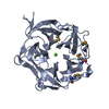
 分子量: 92.094 Da / 分子数: 2 / 由来タイプ: 合成 / 式: C3H8O3
 分子量: 24.305 Da / 分子数: 1 / 由来タイプ: 合成 / 式: Mg / タイプ: SUBJECT OF INVESTIGATION
 分子量: 18.015 Da / 分子数: 463 / 由来タイプ: 天然 / 式: H2O
 試料調製
試料調製 解析
解析