 Movie
Movie Controller

[English] 日本語
 Yorodumi
- PDB-6p6q: HCV NS3/4A protease domain of genotype 1a3a chimera in complex wi... -
+ Open data

- Basic information
| Entry | Database: PDB / ID: 6p6q | ||||||
|---|---|---|---|---|---|---|---|
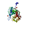
| Title | HCV NS3/4A protease domain of genotype 1a3a chimera in complex with grazoprevir | ||||||
 Components Components | Non-structural protein 4A,Serine protease NS3 | ||||||
 Keywords Keywords | VIRAL PROTEIN / Hydrolase / HCV NS3/4A protease HCV protease domain Grazoprevir / GZR Genotype 1a3a chimera | ||||||
| Function / homology |  Function and homology information Function and homology informationpositive regulation of metabolic process / regulation of primary metabolic process / hepacivirin / host cell mitochondrial membrane / host cell lipid droplet / symbiont-mediated transformation of host cell / symbiont-mediated suppression of host TRAF-mediated signal transduction / symbiont-mediated perturbation of host cell cycle G1/S transition checkpoint / symbiont-mediated suppression of host JAK-STAT cascade via inhibition of STAT1 activity / symbiont-mediated suppression of host cytoplasmic pattern recognition receptor signaling pathway via inhibition of MAVS activity ...positive regulation of metabolic process / regulation of primary metabolic process / hepacivirin / host cell mitochondrial membrane / host cell lipid droplet / symbiont-mediated transformation of host cell / symbiont-mediated suppression of host TRAF-mediated signal transduction / symbiont-mediated perturbation of host cell cycle G1/S transition checkpoint / symbiont-mediated suppression of host JAK-STAT cascade via inhibition of STAT1 activity / symbiont-mediated suppression of host cytoplasmic pattern recognition receptor signaling pathway via inhibition of MAVS activity / SH3 domain binding / nucleoside-triphosphate phosphatase / viral nucleocapsid / channel activity / monoatomic ion transmembrane transport / clathrin-dependent endocytosis of virus by host cell / Hydrolases; Acting on peptide bonds (peptidases); Cysteine endopeptidases / RNA helicase activity / host cell perinuclear region of cytoplasm / host cell endoplasmic reticulum membrane / RNA helicase / symbiont-mediated suppression of host type I interferon-mediated signaling pathway / ribonucleoprotein complex / serine-type endopeptidase activity / viral translational frameshifting / symbiont-mediated activation of host autophagy / RNA-directed RNA polymerase / cysteine-type endopeptidase activity / viral RNA genome replication / RNA-directed RNA polymerase activity / fusion of virus membrane with host endosome membrane / viral envelope / virion attachment to host cell / host cell nucleus / host cell plasma membrane / virion membrane / structural molecule activity / DNA-templated transcription / ATP hydrolysis activity / proteolysis / RNA binding / zinc ion binding / ATP binding Similarity search - Function | ||||||
| Biological species |  Hepatitis C virus genotype 1a Hepatitis C virus genotype 1a | ||||||
| Method |  X-RAY DIFFRACTION / SYNCHROTRON / MOLECULAR REPLACEMENT / Resolution: 3.5 Å X-RAY DIFFRACTION / SYNCHROTRON / MOLECULAR REPLACEMENT / Resolution: 3.5 Å | ||||||
 Authors Authors | Timm, J. / Schiffer, C.A. | ||||||
| Funding support |  United States, 1items United States, 1items
| ||||||
 Citation Citation | Journal: To Be Published Title: Molecular mechanism of pan-genotypic HCV NS3/4A protease inhibition by glecaprevir and characterization of genotype-specific structural differences Authors: Timm, J. / Kosovrasti, K. / Henes, M. / Leidner, F. / Hou, S. / Kurt-Yilmaz, N. / Schiffer, C.A. | ||||||
| History |
|
- Structure visualization
| Structure viewer | Molecule: MolmilJmol/JSmol |
|---|
- Downloads & links
-Download
| PDBx/mmCIF format | 6p6q.cif.gz | 146 KB | Display | PDBx/mmCIF format |
|---|---|---|---|---|
| PDB format | pdb6p6q.ent.gz | 113.1 KB | Display | PDB format |
| PDBx/mmJSON format | 6p6q.json.gz | Tree view | PDBx/mmJSON format | |
| Others |  Other downloads Other downloads |
-Validation report
| Arichive directory | https://data.pdbj.org/pub/pdb/validation_reports/p6/6p6qftp://data.pdbj.org/pub/pdb/validation_reports/p6/6p6q | HTTPS FTP |
|---|
-Related structure data
| Related structure data |  6p6rC  6p6sC  6p6tC  6p6vC  6p6zC  5vojS S: Starting model for refinement C: citing same article ( |
|---|---|
| Similar structure data |


-Links
 PDBj
PDBj
- Assembly
| Deposited unit | 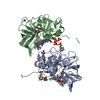
| ||||||||
|---|---|---|---|---|---|---|---|---|---|
| 1 | 
| ||||||||
| 2 | 
| ||||||||
| Unit cell |
|
-Components
-Protein , 1 types, 2 molecules AB
| #1: Protein | Mass: 21460.281 Da / Num. of mol.: 2 Mutation: C991S, V9941, V995N, L1013E, L1014E, I1017Q, I1018E, L1021Q, A1040T, C1047S, C1052L, I1072T, Q1080K, P1086Q, N1174S, C1159S, I1132L, D1168Q, R1123T Source method: isolated from a genetically manipulated source Source: (gene. exp.) Hepatitis C virus genotype 1a (isolate 1)Strain: isolate 1 / Production host:  References: UniProt: P26664, hepacivirin, nucleoside-triphosphate phosphatase, RNA helicase |
|---|
-Non-polymers , 5 types, 65 molecules 


| #2: Chemical |  Mass: 766.903 Da / Num. of mol.: 2 / Source method: obtained synthetically / Formula: C38H50N6O9S / Feature type: SUBJECT OF INVESTIGATION / Comment: protease inhibitor*YM Mass: 766.903 Da / Num. of mol.: 2 / Source method: obtained synthetically / Formula: C38H50N6O9S / Feature type: SUBJECT OF INVESTIGATION / Comment: protease inhibitor*YM#3: Chemical |  Mass: 65.409 Da / Num. of mol.: 2 / Source method: obtained synthetically / Formula: Zn Mass: 65.409 Da / Num. of mol.: 2 / Source method: obtained synthetically / Formula: Zn#4: Chemical |  Mass: 195.237 Da / Num. of mol.: 2 / Source method: obtained synthetically / Formula: C6H13NO4S / Comment: pH buffer*YM Mass: 195.237 Da / Num. of mol.: 2 / Source method: obtained synthetically / Formula: C6H13NO4S / Comment: pH buffer*YM#5: Chemical | ChemComp-SO4 /  Mass: 96.063 Da / Num. of mol.: 7 / Source method: obtained synthetically / Formula: SO4 Mass: 96.063 Da / Num. of mol.: 7 / Source method: obtained synthetically / Formula: SO4#6: Water | ChemComp-HOH / | Mass: 18.015 Da / Num. of mol.: 52 / Source method: isolated from a natural source / Formula: H2O |
|---|
-Experimental details
-Experiment
| Experiment | Method: X-RAY DIFFRACTION / Number of used crystals: 1 |
|---|
- Sample preparation
| Crystal | Density Matthews: 2.22 Å3/Da / Density % sol: 44.49 % |
|---|---|
| Crystal grow | Temperature: 291 K / Method: vapor diffusion Details: 0.1 M MES pH 6.5 5 % (NH4)2SO4 24 % PEG 3350 3 mM grazoprevir |
-Data collection
| Diffraction | Mean temperature: 100 K / Serial crystal experiment: N |
|---|---|
| Diffraction source | Source: SYNCHROTRON / Site: APS / Beamline: 21-ID-D / Wavelength: 1.0332 Å |
| Detector | Type: DECTRIS PILATUS3 6M / Detector: PIXEL / Date: Jun 24, 2018 |
| Radiation | Protocol: SINGLE WAVELENGTH / Monochromatic (M) / Laue (L): M / Scattering type: x-ray |
| Radiation wavelength | Wavelength: 1.0332 Å / Relative weight: 1 |
| Reflection | Resolution: 3.5→48.954 Å / Num. obs: 9272 / % possible obs: 99.86 % / Redundancy: 2 % / Rmerge(I) obs: 0.0819 / Net I/σ(I): 7.18 |
| Reflection shell | Resolution: 3.5→3.625 Å / Rmerge(I) obs: 0.1376 / Num. unique obs: 475 |
- Processing
| Software |
| ||||||||||||||||||||||||||||
|---|---|---|---|---|---|---|---|---|---|---|---|---|---|---|---|---|---|---|---|---|---|---|---|---|---|---|---|---|---|
| Refinement | Method to determine structure: MOLECULAR REPLACEMENT Starting model: 5voj Resolution: 3.5→48.954 Å / SU ML: 0.38 / Cross valid method: FREE R-VALUE / σ(F): 1.36 / Phase error: 26.67
| ||||||||||||||||||||||||||||
| Solvent computation | Shrinkage radii: 0.9 Å / VDW probe radii: 1.11 Å | ||||||||||||||||||||||||||||
| Refinement step | Cycle: LAST / Resolution: 3.5→48.954 Å
| ||||||||||||||||||||||||||||
| Refine LS restraints |
| ||||||||||||||||||||||||||||
| LS refinement shell |
|
