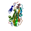
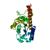
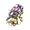
Entry Database : PDB / ID : 5vojTitle Crystal structure of HCV NS3/4A protease in complex with JZ01-15, an analogue of 5172-mcP1P3 NS4A cofactor -- NS3 protein chimera Keywords / / / / / Function / homology Function Domain/homology Component
/ / / / / / / / / / / / / / / / / / / / / / / / / / / / / / / / / / / / / / / / / / / / / / / / / / / / / / / / / / / / / / / / / / / / / / / / / / / / / / / / / / / / / / / / / / / / / / / / / / / / / / / / / / / / / / / / / / / / Biological species Method / / Resolution : 1.799 Å Authors Matthew, A.N. / Schiffer, C.A. Funding support Organization Grant number Country National Institutes of Health/National Institute Of Allergy and Infectious Diseases (NIH/NIAID) R01 AI085051 National Institutes of Health/National Institute of General Medical Sciences (NIH/NIGMS) F31 GM119345
Journal : J. Med. Chem. / Year : 2017Title : Hepatitis C Virus NS3/4A Protease Inhibitors Incorporating Flexible P2 Quinoxalines Target Drug Resistant Viral Variants.Authors : Matthew, A.N. / Zephyr, J. / Hill, C.J. / Jahangir, M. / Newton, A. / Petropoulos, C.J. / Huang, W. / Kurt-Yilmaz, N. / Schiffer, C.A. / Ali, A. History Deposition May 3, 2017 Deposition site / Processing site Revision 1.0 Jun 21, 2017 Provider / Type Revision 1.1 Jul 26, 2017 Group / Category / citation_authorItem _citation.journal_volume / _citation.page_first ... _citation.journal_volume / _citation.page_first / _citation.page_last / _citation_author.name Revision 1.2 Sep 20, 2017 Group / Category / Item Revision 1.3 Dec 11, 2019 Group / Category / Item Revision 1.4 Oct 4, 2023 Group / Database references / Refinement descriptionCategory chem_comp_atom / chem_comp_bond ... chem_comp_atom / chem_comp_bond / database_2 / pdbx_initial_refinement_model Item / _database_2.pdbx_database_accession
Show all Show less
 Movie
Movie Controller

 Yorodumi
 Open data
 Basic information
 Components
 Keywords
 Function and homology information
 Hepatitis C virus subtype 1a
 X-RAY DIFFRACTION /
  Authors
 United States, 2items
  Citation
 Structure visualization
 Downloads & links
 Other downloads


 PDBj
PDBj
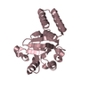
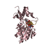
 Assembly

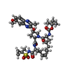
 Mass: 754.893 Da / Num. of mol.: 1 / Source method: obtained synthetically / Formula: C37H50N6O9S
 Mass: 96.063 Da / Num. of mol.: 1 / Source method: obtained synthetically / Formula: SO4
 Mass: 65.409 Da / Num. of mol.: 1 / Source method: obtained synthetically / Formula: Zn
 Mass: 18.015 Da / Num. of mol.: 183 / Source method: isolated from a natural source / Formula: H2O
 Sample preparation
 Processing