Deposited unit
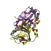
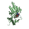
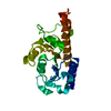
A: Cupin 2, conserved barrel domain protein
B: Cupin 2, conserved barrel domain protein
C: Cupin 2, conserved barrel domain protein
D: Cupin 2, conserved barrel domain protein
E: Cupin 2, conserved barrel domain protein
hetero molecules Summary Component details
Theoretical mass Number of molelcules Total (without water) 61,889 17 Polymers 61,411 5 Non-polymers 478 12 Water 4,270 237
1
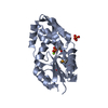
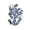
D: Cupin 2, conserved barrel domain protein
E: Cupin 2, conserved barrel domain protein
hetero molecules Summary Component details Symmetry operations Calculated values
Theoretical mass Number of molelcules Total (without water) 24,866 8 Polymers 24,564 2 Non-polymers 302 6 Water 36 2
Type Name Symmetry operation Number identity operation 1_555 x,y,z 1
Buried area 4470 Å2 ΔGint -54 kcal/mol Surface area 9610 Å2 Method
2
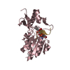
A: Cupin 2, conserved barrel domain protein
hetero molecules
A: Cupin 2, conserved barrel domain protein
hetero molecules Summary Component details Symmetry operations Calculated values
Theoretical mass Number of molelcules Total (without water) 24,682 6 Polymers 24,564 2 Non-polymers 117 4 Water 36 2
Type Name Symmetry operation Number identity operation 1_555 x,y,z 1 crystal symmetry operation 4_565 x,-y+1,-z 1
Buried area 3810 Å2 ΔGint -52 kcal/mol Surface area 9940 Å2 Method
3
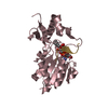
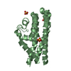
B: Cupin 2, conserved barrel domain protein
C: Cupin 2, conserved barrel domain protein
hetero molecules Summary Component details Symmetry operations Calculated values
Theoretical mass Number of molelcules Total (without water) 24,682 6 Polymers 24,564 2 Non-polymers 117 4 Water 36 2
Type Name Symmetry operation Number identity operation 1_555 x,y,z 1
Buried area 3280 Å2 ΔGint -48 kcal/mol Surface area 9810 Å2 Method
Unit cell Length a, b, c (Å) 56.920, 95.140, 237.370 Angle α, β, γ (deg.) 90.000, 90.000, 90.000 Int Tables number 20 Space group name H-M C2221
Components on special symmetry positions ID Model Components 1 1 E -554-HOH
Noncrystallographic symmetry (NCS) NCS domain ID Ens-ID Details (eV)1 1 A2 1 B3 1 C4 1 D5 1 E6 1 A7 1 B8 1 C9 1 D10 1 E11 1 A12 1 B13 1 C14 1 D15 1 E
NCS domain segments Ens-ID
Dom-ID Component-ID Beg auth comp-ID Beg label comp-ID End auth comp-ID End label comp-ID Refine code Auth asym-ID Label asym-ID Auth seq-ID Label seq-ID 1 1 VALVALPHEPHE4 AA5 - 64 6 - 65 2 1 VALVALPHEPHE4 BB5 - 64 6 - 65 3 1 VALVALPHEPHE4 CC5 - 64 6 - 65 4 1 VALVALPHEPHE4 DD5 - 64 6 - 65 5 1 VALVALPHEPHE4 EE5 - 64 6 - 65 6 2 ARGARGASPASP4 AA65 - 66 66 - 67 7 2 ARGARGASPASP4 BB65 - 66 66 - 67 8 2 ARGARGASPASP4 CC65 - 66 66 - 67 9 2 ARGARGASPASP4 DD65 - 66 66 - 67 10 2 ARGARGASPASP4 EE65 - 66 66 - 67 11 3 GLNGLNARGARG6 AA67 - 101 68 - 102 12 3 GLNGLNARGARG6 BB67 - 101 68 - 102 13 3 GLNGLNARGARG6 CC
 Movie
Movie Controller

 Yorodumi
 Open data
 Basic information
 Components
 Keywords
 Function and homology information
 Shewanella frigidimarina NCIMB 400 (bacteria)
 X-RAY DIFFRACTION /
  Authors
 Citation
 Structure visualization
 Downloads & links
 Other downloads
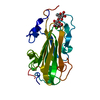


 PDBj
PDBj

 Assembly