 Movie
Movie Controller

[English] 日本語
 Yorodumi
- PDB-6p58: Dark and Steady State-Illuminated Crystal Structure of Cyanobacte... -
+ Open data

- Basic information
| Entry | Database: PDB / ID: 6p58 | |||||||||
|---|---|---|---|---|---|---|---|---|---|---|
| Title | Dark and Steady State-Illuminated Crystal Structure of Cyanobacteriochrome Receptor PixJ at 150K | |||||||||
 Components Components | Methyl-accepting chemotaxis protein | |||||||||
 Keywords Keywords | SIGNALING PROTEIN / phytochrome / bilin / light-sensing / temperature-scan | |||||||||
| Function / homology |  Function and homology information Function and homology information | |||||||||
| Biological species |   Thermosynechococcus elongatus (bacteria) Thermosynechococcus elongatus (bacteria) | |||||||||
| Method |  X-RAY DIFFRACTION / SYNCHROTRON / MOLECULAR REPLACEMENT / Resolution: 1.499 Å X-RAY DIFFRACTION / SYNCHROTRON / MOLECULAR REPLACEMENT / Resolution: 1.499 Å | |||||||||
 Authors Authors | Clinger, J.A. / Miller, M.D. / Buirgie, E.S. / Vierstra, R.D. / Phillips Jr., G.N. | |||||||||
| Funding support |  United States, 2items United States, 2items
| |||||||||
 Citation Citation | Journal: Proc.Natl.Acad.Sci.USA / Year: 2020 Title: Photoreversible interconversion of a phytochrome photosensory module in the crystalline state. Authors: Burgie, E.S. / Clinger, J.A. / Miller, M.D. / Brewster, A.S. / Aller, P. / Butryn, A. / Fuller, F.D. / Gul, S. / Young, I.D. / Pham, C.C. / Kim, I.S. / Bhowmick, A. / O'Riordan, L.J. / ...Authors: Burgie, E.S. / Clinger, J.A. / Miller, M.D. / Brewster, A.S. / Aller, P. / Butryn, A. / Fuller, F.D. / Gul, S. / Young, I.D. / Pham, C.C. / Kim, I.S. / Bhowmick, A. / O'Riordan, L.J. / Sutherlin, K.D. / Heinemann, J.V. / Batyuk, A. / Alonso-Mori, R. / Hunter, M.S. / Koglin, J.E. / Yano, J. / Yachandra, V.K. / Sauter, N.K. / Cohen, A.E. / Kern, J. / Orville, A.M. / Phillips Jr., G.N. / Vierstra, R.D. | |||||||||
| History |
|
- Structure visualization
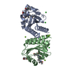
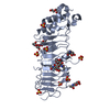
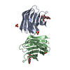
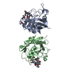
| Structure viewer | Molecule: MolmilJmol/JSmol |
|---|
- Downloads & links
-Download
| PDBx/mmCIF format | 6p58.cif.gz | 213 KB | Display | PDBx/mmCIF format |
|---|---|---|---|---|
| PDB format | pdb6p58.ent.gz | 173 KB | Display | PDB format |
| PDBx/mmJSON format | 6p58.json.gz | Tree view | PDBx/mmJSON format | |
| Others |  Other downloads Other downloads |
-Validation report
| Arichive directory | https://data.pdbj.org/pub/pdb/validation_reports/p5/6p58ftp://data.pdbj.org/pub/pdb/validation_reports/p5/6p58 | HTTPS FTP |
|---|
-Related structure data
| Related structure data |  6pruC  6pryC  6uppC  4glqS S: Starting model for refinement C: citing same article ( |
|---|---|
| Similar structure data |


-Links
 PDBj
PDBj


- Assembly
| Deposited unit | 
| ||||||||
|---|---|---|---|---|---|---|---|---|---|
| 1 |
| ||||||||
| Unit cell |
|
-Components
| #1: Protein | Mass: 17152.381 Da / Num. of mol.: 2 / Fragment: GAF domain / Mutation: C555A Source method: isolated from a genetically manipulated source Source: (gene. exp.) Thermosynechococcus elongatus (strain BP-1) (bacteria)Strain: BP-1 / Gene: tll0569 / Production host: #2: Chemical |   Mass: 590.710 Da / Num. of mol.: 2 / Source method: obtained synthetically / Formula: C33H42N4O6 / Feature type: SUBJECT OF INVESTIGATION Mass: 590.710 Da / Num. of mol.: 2 / Source method: obtained synthetically / Formula: C33H42N4O6 / Feature type: SUBJECT OF INVESTIGATION#3: Chemical | ChemComp-MG /   Mass: 24.305 Da / Num. of mol.: 4 / Source method: obtained synthetically / Formula: Mg Mass: 24.305 Da / Num. of mol.: 4 / Source method: obtained synthetically / Formula: Mg#4: Chemical |   Mass: 62.068 Da / Num. of mol.: 2 / Source method: obtained synthetically / Formula: C2H6O2 Mass: 62.068 Da / Num. of mol.: 2 / Source method: obtained synthetically / Formula: C2H6O2#5: Water | ChemComp-HOH / |  Mass: 18.015 Da / Num. of mol.: 293 / Source method: isolated from a natural source / Formula: H2O Mass: 18.015 Da / Num. of mol.: 293 / Source method: isolated from a natural source / Formula: H2OHas protein modification | Y | |
|---|
-Experimental details
-Experiment
| Experiment | Method: X-RAY DIFFRACTION / Number of used crystals: 1 |
|---|
- Sample preparation
| Crystal | Density Matthews: 2.15 Å3/Da / Density % sol: 42.81 % |
|---|---|
| Crystal grow | Temperature: 298 K / Method: liquid diffusion / pH: 7.5 / Details: 100mM HEPES pH 7.5, 18% w/v PEG 3350, 200mM MgCl2 |
-Data collection
| Diffraction | Mean temperature: 150 K / Serial crystal experiment: N | ||||||||||||||||||||||||||||||
|---|---|---|---|---|---|---|---|---|---|---|---|---|---|---|---|---|---|---|---|---|---|---|---|---|---|---|---|---|---|---|---|
| Diffraction source | Source: SYNCHROTRON / Site: APS / Beamline: 21-ID-D / Wavelength: 0.9919 Å | ||||||||||||||||||||||||||||||
| Detector | Type: DECTRIS EIGER X 16M / Detector: PIXEL / Date: Mar 24, 2019 | ||||||||||||||||||||||||||||||
| Radiation | Protocol: SINGLE WAVELENGTH / Monochromatic (M) / Laue (L): M / Scattering type: x-ray | ||||||||||||||||||||||||||||||
| Radiation wavelength | Wavelength: 0.9919 Å / Relative weight: 1 | ||||||||||||||||||||||||||||||
| Reflection | Resolution: 1.5→33.8 Å / Num. obs: 48186 / % possible obs: 99.8 % / Redundancy: 6.6 % / CC1/2: 0.998 / Rmerge(I) obs: 0.113 / Rpim(I) all: 0.047 / Rrim(I) all: 0.123 / Net I/σ(I): 8 / Num. measured all: 320193 / Scaling rejects: 348 | ||||||||||||||||||||||||||||||
| Reflection shell | Diffraction-ID: 1
|
- Processing
| Software |
| ||||||||||||||||||||||||||||||||||||||||||||||||||||||||||||||||||||||||||||||||||||||||||||||||||||||||||||||||||||||||||||||
|---|---|---|---|---|---|---|---|---|---|---|---|---|---|---|---|---|---|---|---|---|---|---|---|---|---|---|---|---|---|---|---|---|---|---|---|---|---|---|---|---|---|---|---|---|---|---|---|---|---|---|---|---|---|---|---|---|---|---|---|---|---|---|---|---|---|---|---|---|---|---|---|---|---|---|---|---|---|---|---|---|---|---|---|---|---|---|---|---|---|---|---|---|---|---|---|---|---|---|---|---|---|---|---|---|---|---|---|---|---|---|---|---|---|---|---|---|---|---|---|---|---|---|---|---|---|---|---|
| Refinement | Method to determine structure: MOLECULAR REPLACEMENT Starting model: 4GLQ Resolution: 1.499→33.828 Å / SU ML: 0.18 / Cross valid method: THROUGHOUT / σ(F): 1.33 / Phase error: 19.27
| ||||||||||||||||||||||||||||||||||||||||||||||||||||||||||||||||||||||||||||||||||||||||||||||||||||||||||||||||||||||||||||||
| Solvent computation | Shrinkage radii: 0.9 Å / VDW probe radii: 1.11 Å | ||||||||||||||||||||||||||||||||||||||||||||||||||||||||||||||||||||||||||||||||||||||||||||||||||||||||||||||||||||||||||||||
| Displacement parameters | Biso max: 148.42 Å2 / Biso mean: 25.9884 Å2 / Biso min: 10.72 Å2 | ||||||||||||||||||||||||||||||||||||||||||||||||||||||||||||||||||||||||||||||||||||||||||||||||||||||||||||||||||||||||||||||
| Refinement step | Cycle: final / Resolution: 1.499→33.828 Å
| ||||||||||||||||||||||||||||||||||||||||||||||||||||||||||||||||||||||||||||||||||||||||||||||||||||||||||||||||||||||||||||||
| LS refinement shell | Refine-ID: X-RAY DIFFRACTION / Rfactor Rfree error: 0 / Total num. of bins used: 17
|
