 Movie
Movie Controller

[English] 日本語
 Yorodumi
- PDB-2pwj: Structure of a mitochondrial type II peroxiredoxin from Pisum sativum -
+ Open data

- Basic information
| Entry | Database: PDB / ID: 2pwj | ||||||
|---|---|---|---|---|---|---|---|
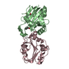
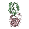
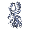
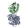
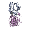
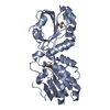
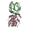
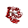
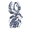
| Title | Structure of a mitochondrial type II peroxiredoxin from Pisum sativum | ||||||
 Components Components | Mitochondrial peroxiredoxin | ||||||
 Keywords Keywords | OXIDOREDUCTASE / alpha and beta protein | ||||||
| Function / homology |  Function and homology information Function and homology informationglutaredoxin-dependent peroxiredoxin / thioredoxin peroxidase activity / cell redox homeostasis / hydrogen peroxide catabolic process / cellular response to oxidative stress / mitochondrial matrix Similarity search - Function | ||||||
| Biological species |  Pisum sativum (garden pea) Pisum sativum (garden pea) | ||||||
| Method |  X-RAY DIFFRACTION / MOLECULAR REPLACEMENT / Resolution: 2.8 Å X-RAY DIFFRACTION / MOLECULAR REPLACEMENT / Resolution: 2.8 Å | ||||||
 Authors Authors | Lopez-Jaramillo, F.J. / Barranco-Medina, S. / Lazaro, J.J. / Santoyo-Gonzalez, F. | ||||||
 Citation Citation | Journal: To be Published Title: Structure of a mitochondrial type II peroxiredoxin from Pisum sativum Authors: Lopez-Jaramillo, F.J. / Barranco-Medina, S. / Lazaro, J.J. / Santoyo-Gonzalez, F. #1: Journal: Acta Crystallogr.,Sect.F / Year: 2006Title: Cloning, overexpression, purification and preliminary crystallographic studies of a mitochondrial type II peroxiredoxin from Pisum sativum Authors: Barranco-Medina, S. / Lopez-Jaramillo, F.J. / Bernier-Villamor, L. / Sevilla, F. / Lazaro, J.J. | ||||||
| History |
|
- Structure visualization
| Structure viewer | Molecule: MolmilJmol/JSmol |
|---|
- Downloads & links
-Download
| PDBx/mmCIF format | 2pwj.cif.gz | 186.4 KB | Display | PDBx/mmCIF format |
|---|---|---|---|---|
| PDB format | pdb2pwj.ent.gz | 150.8 KB | Display | PDB format |
| PDBx/mmJSON format | 2pwj.json.gz | Tree view | PDBx/mmJSON format | |
| Others |  Other downloads Other downloads |
-Validation report
| Arichive directory | https://data.pdbj.org/pub/pdb/validation_reports/pw/2pwjftp://data.pdbj.org/pub/pdb/validation_reports/pw/2pwj | HTTPS FTP |
|---|
-Related structure data
| Similar structure data |
|---|

-Links
 PDBj
PDBj- Assembly
| Deposited unit | 
| ||||||||
|---|---|---|---|---|---|---|---|---|---|
| 1 | 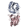
| ||||||||
| 2 | 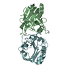
| ||||||||
| 3 | 
| ||||||||
| Unit cell |
| ||||||||
| Details | The biological assembly seems to be a dimer althoug hexameric forms have been detected. This point is beind elucidated by biochemical experiments and analysis of this structure |
-Components
| #1: Protein | Mass: 18464.836 Da / Num. of mol.: 6 Source method: isolated from a genetically manipulated source Source: (gene. exp.) Pisum sativum (garden pea) / Gene: prx / Plasmid: pET3d_Prx / Production host:  Has protein modification | N | |
|---|
-Experimental details
-Experiment
| Experiment | Method: X-RAY DIFFRACTION / Number of used crystals: 1 |
|---|
- Sample preparation
| Crystal | Density Matthews: 2.74 Å3/Da / Density % sol: 55.16 % |
|---|---|
| Crystal grow | Temperature: 293 K / Method: vapor diffusion, hanging drop / pH: 5.6 Details: PEG2000, citrate, NaCl, DTT, 2-propanol, pH 5.6, VAPOR DIFFUSION, HANGING DROP, temperature 293K |
-Data collection
| Diffraction | Mean temperature: 100 K | |||||||||||||||||||||||||||||||||
|---|---|---|---|---|---|---|---|---|---|---|---|---|---|---|---|---|---|---|---|---|---|---|---|---|---|---|---|---|---|---|---|---|---|---|
| Diffraction source | Source: ROTATING ANODE / Type: BRUKER AXS MICROSTAR-H / Wavelength: 1.5418 | |||||||||||||||||||||||||||||||||
| Detector | Type: BRUKER PLATINUM 200 / Detector: CCD / Date: Feb 2, 2005 / Details: Microstar micro-focus | |||||||||||||||||||||||||||||||||
| Radiation | Monochromator: mirrors / Protocol: SINGLE WAVELENGTH / Monochromatic (M) / Laue (L): M / Scattering type: x-ray | |||||||||||||||||||||||||||||||||
| Radiation wavelength | Wavelength: 1.5418 Å / Relative weight: 1 | |||||||||||||||||||||||||||||||||
| Reflection | Resolution: 2.39→71.9 Å / Num. all: 30073 / Num. obs: 30073 / % possible obs: 65.79 % / Observed criterion σ(F): 0 / Observed criterion σ(I): 0 / Redundancy: 3.08 % / Biso Wilson estimate: 22.1 Å2 / Limit h max: 20 / Limit h min: -24 / Limit k max: 25 / Limit k min: -24 / Limit l max: 31 / Limit l min: 0 / Observed criterion F max: 1058698.45 / Observed criterion F min: 1.31 / Rsym value: 0.067 / Net I/σ(I): 15.416 | |||||||||||||||||||||||||||||||||
| Reflection shell |
|
-Phasing
| Phasing MR | Rfactor: 0.613 / Cor.coef. Fo:Fc: 0.3 / Cor.coef. Io to Ic: 0.231
|
|---|
- Processing
| Software |
| ||||||||||||||||||||||||||||||||||||||||||||||||||||||||||||||||||||||||||||||||||||||||||
|---|---|---|---|---|---|---|---|---|---|---|---|---|---|---|---|---|---|---|---|---|---|---|---|---|---|---|---|---|---|---|---|---|---|---|---|---|---|---|---|---|---|---|---|---|---|---|---|---|---|---|---|---|---|---|---|---|---|---|---|---|---|---|---|---|---|---|---|---|---|---|---|---|---|---|---|---|---|---|---|---|---|---|---|---|---|---|---|---|---|---|---|
| Refinement | Method to determine structure: MOLECULAR REPLACEMENT / Resolution: 2.8→14.98 Å / Rfactor Rfree error: 0.006 / Occupancy max: 1 / Occupancy min: 1 / Cross valid method: THROUGHOUT / σ(F): 0 / Stereochemistry target values: Engh & Huber Details: Cross-validation method: -> "throughout" Free R value test set selection criteria: -> "random" Number of non-hydrogen atoms used in refinement. Polymer 7476 Nonpolymer 0 Solvent 0 CNS ...Details: Cross-validation method: -> "throughout" Free R value test set selection criteria: -> "random" Number of non-hydrogen atoms used in refinement. Polymer 7476 Nonpolymer 0 Solvent 0 CNS Parameter files: CNS_TOPPAR/protein_rep.param CNS_TOPPAR/carbohydrate.param CNS Topology files: CNS_TOPPAR/protein.top CNS_TOPPAR/carbohydrate.top
| ||||||||||||||||||||||||||||||||||||||||||||||||||||||||||||||||||||||||||||||||||||||||||
| Solvent computation | Solvent model: CNS bulk solvent model used / Bsol: 14.9103 Å2 / ksol: 0.296614 e/Å3 | ||||||||||||||||||||||||||||||||||||||||||||||||||||||||||||||||||||||||||||||||||||||||||
| Displacement parameters | Biso max: 39.46 Å2 / Biso mean: 14.29 Å2 / Biso min: 5.4 Å2
| ||||||||||||||||||||||||||||||||||||||||||||||||||||||||||||||||||||||||||||||||||||||||||
| Refine analyze |
| ||||||||||||||||||||||||||||||||||||||||||||||||||||||||||||||||||||||||||||||||||||||||||
| Refinement step | Cycle: LAST / Resolution: 2.8→14.98 Å
| ||||||||||||||||||||||||||||||||||||||||||||||||||||||||||||||||||||||||||||||||||||||||||
| Refine LS restraints |
| ||||||||||||||||||||||||||||||||||||||||||||||||||||||||||||||||||||||||||||||||||||||||||
| LS refinement shell | Refine-ID: X-RAY DIFFRACTION
|
