 Movie
Movie Controller


+ Open data

- Basic information
| Entry | Database: PDB / ID: 4k7n | ||||||
|---|---|---|---|---|---|---|---|
| Title | HUMAN PEROXIREDOXIN 5 with a fragment | ||||||
 Components Components | Peroxiredoxin-5, mitochondrial | ||||||
 Keywords Keywords | OXIDOREDUCTASE / enzyme / cytosol | ||||||
| Function / homology |  Function and homology information Function and homology informationperoxynitrite reductase activity / reactive nitrogen species metabolic process / negative regulation of transcription by RNA polymerase III / negative regulation of oxidoreductase activity / regulation of apoptosis involved in tissue homeostasis / RNA polymerase III transcription regulatory region sequence-specific DNA binding / thioredoxin-dependent peroxiredoxin / thioredoxin peroxidase activity / cysteine-type endopeptidase inhibitor activity involved in apoptotic process / Detoxification of Reactive Oxygen Species ...peroxynitrite reductase activity / reactive nitrogen species metabolic process / negative regulation of transcription by RNA polymerase III / negative regulation of oxidoreductase activity / regulation of apoptosis involved in tissue homeostasis / RNA polymerase III transcription regulatory region sequence-specific DNA binding / thioredoxin-dependent peroxiredoxin / thioredoxin peroxidase activity / cysteine-type endopeptidase inhibitor activity involved in apoptotic process / Detoxification of Reactive Oxygen Species / antioxidant activity / peroxisomal matrix / positive regulation of collagen biosynthetic process / intracellular membrane-bounded organelle / cell redox homeostasis / hydrogen peroxide catabolic process / TP53 Regulates Metabolic Genes / peroxidase activity / cellular response to reactive oxygen species / peroxisome / cellular response to oxidative stress / response to oxidative stress / cytoplasmic vesicle / mitochondrial matrix / inflammatory response / signaling receptor binding / negative regulation of apoptotic process / perinuclear region of cytoplasm / mitochondrion / : / extracellular exosome / identical protein binding / nucleus / cytoplasm / cytosol Similarity search - Function | ||||||
| Biological species |  Homo sapiens (human) Homo sapiens (human) | ||||||
| Method |  X-RAY DIFFRACTION / SYNCHROTRON / MOLECULAR REPLACEMENT / Resolution: 2.3 Å X-RAY DIFFRACTION / SYNCHROTRON / MOLECULAR REPLACEMENT / Resolution: 2.3 Å | ||||||
 Authors Authors | Guichou, J.F. | ||||||
 Citation Citation | Journal: Plos One / Year: 2014 Title: Comparing Binding Modes of Analogous Fragments Using NMR in Fragment-Based Drug Design: Application to PRDX5 Authors: Aguirre, C. / Brink, T.T. / Guichou, J.F. / Cala, O. / Krimm, I. | ||||||
| History |
|
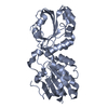
- Structure visualization
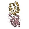
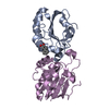
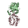
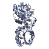
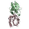
| Structure viewer | Molecule: MolmilJmol/JSmol |
|---|
- Downloads & links
-Download
| PDBx/mmCIF format | 4k7n.cif.gz | 104 KB | Display | PDBx/mmCIF format |
|---|---|---|---|---|
| PDB format | pdb4k7n.ent.gz | 80.8 KB | Display | PDB format |
| PDBx/mmJSON format | 4k7n.json.gz | Tree view | PDBx/mmJSON format | |
| Others |  Other downloads Other downloads |
-Validation report
| Arichive directory | https://data.pdbj.org/pub/pdb/validation_reports/k7/4k7nftp://data.pdbj.org/pub/pdb/validation_reports/k7/4k7n | HTTPS FTP |
|---|
-Related structure data
| Related structure data |  4k7iC  4k7oC  4mmmC 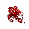 1hd2S C: citing same article ( S: Starting model for refinement |
|---|---|
| Similar structure data |


-Links
 PDBj
PDBj


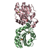
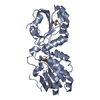
- Assembly
| Deposited unit | 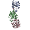
| ||||||||
|---|---|---|---|---|---|---|---|---|---|
| 1 | 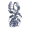
| ||||||||
| 2 | 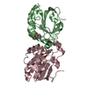
| ||||||||
| Unit cell |
|
-Components
| #1: Protein | Mass: 17835.475 Da / Num. of mol.: 3 / Fragment: UNP residues 54-214 Source method: isolated from a genetically manipulated source Source: (gene. exp.) Homo sapiens (human) / Gene: PRDX5, ACR1, SBBI10 / Production host:  #2: Chemical | 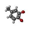  Mass: 124.137 Da / Num. of mol.: 3 / Source method: obtained synthetically / Formula: C7H8O2 Mass: 124.137 Da / Num. of mol.: 3 / Source method: obtained synthetically / Formula: C7H8O2#3: Water | ChemComp-HOH / |  Mass: 18.015 Da / Num. of mol.: 146 / Source method: isolated from a natural source / Formula: H2O Mass: 18.015 Da / Num. of mol.: 146 / Source method: isolated from a natural source / Formula: H2O |
|---|
-Experimental details
-Experiment
| Experiment | Method: X-RAY DIFFRACTION / Number of used crystals: 1 |
|---|
- Sample preparation
| Crystal | Density Matthews: 2.96 Å3/Da / Density % sol: 58.39 % |
|---|---|
| Crystal grow | Temperature: 291 K / Method: vapor diffusion, hanging drop / pH: 5.3 Details: 22% PEG3350, 0.1M sodium citrate buffer, 0.2M potassium sodium tartrate, 5mM 1,4-dithio-dl-threitol, 0.02%(w/v) sodium azide, pH 5.3, VAPOR DIFFUSION, HANGING DROP, temperature 291K |
-Data collection
| Diffraction | Mean temperature: 100 K |
|---|---|
| Diffraction source | Source: SYNCHROTRON / Site: ESRF  / Beamline: ID23-1 / Wavelength: 0.977 Å / Beamline: ID23-1 / Wavelength: 0.977 Å |
| Detector | Type: PSI PILATUS 6M / Detector: PIXEL / Date: Mar 9, 2013 |
| Radiation | Monochromator: graphite / Protocol: SINGLE WAVELENGTH / Monochromatic (M) / Laue (L): M / Scattering type: x-ray |
| Radiation wavelength | Wavelength: 0.977 Å / Relative weight: 1 |
| Reflection | Resolution: 2.3→48.9 Å / Num. all: 27075 / Num. obs: 26994 / % possible obs: 99.7 % / Observed criterion σ(F): 9.1 / Observed criterion σ(I): 1.9 / Redundancy: 5.7 % / Rmerge(I) obs: 0.057 |
| Reflection shell | Resolution: 2.3→48.9 Å / Redundancy: 5.7 % / Mean I/σ(I) obs: 9.1 / % possible all: 99.7 |
- Processing
| Software |
| |||||||||||||||||||||||||
|---|---|---|---|---|---|---|---|---|---|---|---|---|---|---|---|---|---|---|---|---|---|---|---|---|---|---|
| Refinement | Method to determine structure: MOLECULAR REPLACEMENT Starting model: 1HD2 Resolution: 2.3→48.9 Å / Cross valid method: THROUGHOUT / σ(F): 9.1 / Stereochemistry target values: Engh & Huber / Details: HYDROGENS HAVE BEEN ADDED IN THE RIDING POSITIONS
| |||||||||||||||||||||||||
| Displacement parameters | Biso mean: 45.893 Å2
| |||||||||||||||||||||||||
| Refinement step | Cycle: LAST / Resolution: 2.3→48.9 Å
|
