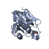
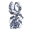
 Movie
Movie Controller


+ Open data

- Basic information
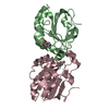
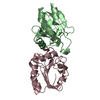
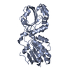
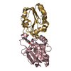
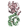
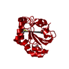
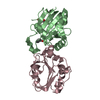
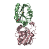
| Entry | Database: PDB / ID: 1hd2 | ||||||
|---|---|---|---|---|---|---|---|
| Title | Human peroxiredoxin 5 | ||||||
 Components Components | PEROXIREDOXIN 5 RESIDUES 54-214 | ||||||
 Keywords Keywords | ANTIOXIDANT ENZYME / PEROXIREDOXIN / THIOREDOXIN PEROXIDASE / THIOREDOXIN FOLD | ||||||
| Function / homology |  Function and homology information Function and homology informationperoxynitrite reductase activity / reactive nitrogen species metabolic process / negative regulation of transcription by RNA polymerase III / negative regulation of oxidoreductase activity / regulation of apoptosis involved in tissue homeostasis / RNA polymerase III transcription regulatory region sequence-specific DNA binding / thioredoxin-dependent peroxiredoxin / thioredoxin peroxidase activity / cysteine-type endopeptidase inhibitor activity involved in apoptotic process / Detoxification of Reactive Oxygen Species ...peroxynitrite reductase activity / reactive nitrogen species metabolic process / negative regulation of transcription by RNA polymerase III / negative regulation of oxidoreductase activity / regulation of apoptosis involved in tissue homeostasis / RNA polymerase III transcription regulatory region sequence-specific DNA binding / thioredoxin-dependent peroxiredoxin / thioredoxin peroxidase activity / cysteine-type endopeptidase inhibitor activity involved in apoptotic process / Detoxification of Reactive Oxygen Species / antioxidant activity / peroxisomal matrix / positive regulation of collagen biosynthetic process / intracellular membrane-bounded organelle / cell redox homeostasis / hydrogen peroxide catabolic process / TP53 Regulates Metabolic Genes / peroxidase activity / cellular response to reactive oxygen species / peroxisome / cellular response to oxidative stress / response to oxidative stress / cytoplasmic vesicle / mitochondrial matrix / inflammatory response / signaling receptor binding / negative regulation of apoptotic process / perinuclear region of cytoplasm / mitochondrion / : / extracellular exosome / identical protein binding / nucleus / cytoplasm / cytosol Similarity search - Function | ||||||
| Biological species |  HOMO SAPIENS (human) HOMO SAPIENS (human) | ||||||
| Method |  X-RAY DIFFRACTION / SYNCHROTRON / MAD / Resolution: 1.5 Å X-RAY DIFFRACTION / SYNCHROTRON / MAD / Resolution: 1.5 Å | ||||||
 Authors Authors | Declercq, J.P. / Evrard, C. | ||||||
 Citation Citation | Journal: J.Mol.Biol. / Year: 2001 Title: Crystal Structure of Human Peroxiredoxin 5, a Novel Type of Mammalian Peroxiredoxin at 1.5 A Resolution. Authors: Declercq, J.P. / Evrard, C. / Clippe, A. / Stricht, D.V. / Bernard, A. / Knoops, B. #1: Journal: J.Biol.Chem. / Year: 1999 Title: Cloning and Characterization of Aoeb166, a Novel Mammalian Antioxidant Enzyme of the Peroxiredoxin Family Authors: Knoops, B. / Clippe, A. / Bogard, C. / Arsalane, K. / Wattiez, R. / Hermans, C. / Duconseille, E. / Falmagne, P. / Bernard, A. | ||||||
| History |
| ||||||
| Remark 650 | HELIX DETERMINATION METHOD: KABSCH AND SANDER | ||||||
| Remark 700 | SHEET DETERMINATION METHOD: KABSCH AND SANDER |
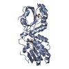
- Structure visualization
| Structure viewer | Molecule: MolmilJmol/JSmol |
|---|
- Downloads & links
-Download
| PDBx/mmCIF format | 1hd2.cif.gz | 82.7 KB | Display | PDBx/mmCIF format |
|---|---|---|---|---|
| PDB format | pdb1hd2.ent.gz | 62.7 KB | Display | PDB format |
| PDBx/mmJSON format | 1hd2.json.gz | Tree view | PDBx/mmJSON format | |
| Others |  Other downloads Other downloads |
-Validation report
| Arichive directory | https://data.pdbj.org/pub/pdb/validation_reports/hd/1hd2ftp://data.pdbj.org/pub/pdb/validation_reports/hd/1hd2 | HTTPS FTP |
|---|
-Related structure data

-Links
 PDBj
PDBj


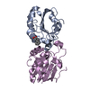
- Assembly
| Deposited unit | 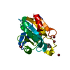
| ||||||||
|---|---|---|---|---|---|---|---|---|---|
| 1 | 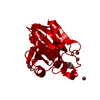
| ||||||||
| Unit cell |
| ||||||||
| Components on special symmetry positions |
|
-Components
| #1: Protein | Mass: 16919.514 Da / Num. of mol.: 1 / Fragment: RESIDUES 54-214 Source method: isolated from a genetically manipulated source Source: (gene. exp.) HOMO SAPIENS (human) / Cellular location: MITOCHONDRIA-CYTOSOL-PEROXISOME / Gene: PRDX5, AOEB166, PMP20, ARC1 / Organ: LUNG / Organelle: MITOCHONDRIA-CYTOSOL-PEROXISOME / Plasmid: PQE-30 / Production host:  | ||
|---|---|---|---|
| #2: Chemical | ChemComp-BEZ /   Mass: 122.121 Da / Num. of mol.: 1 / Source method: obtained synthetically / Formula: C7H6O2 Mass: 122.121 Da / Num. of mol.: 1 / Source method: obtained synthetically / Formula: C7H6O2 | ||
| #3: Chemical | ChemComp-BR /   Mass: 79.904 Da / Num. of mol.: 5 / Source method: obtained synthetically / Formula: Br Mass: 79.904 Da / Num. of mol.: 5 / Source method: obtained synthetically / Formula: Br#4: Water | ChemComp-HOH / |  Mass: 18.015 Da / Num. of mol.: 221 / Source method: isolated from a natural source / Formula: H2O Mass: 18.015 Da / Num. of mol.: 221 / Source method: isolated from a natural source / Formula: H2O |
-Experimental details
-Experiment
| Experiment | Method: X-RAY DIFFRACTION / Number of used crystals: 1 |
|---|
- Sample preparation
| Crystal | Density Matthews: 3.37 Å3/Da / Density % sol: 63.5 % | |||||||||||||||||||||||||||||||||||
|---|---|---|---|---|---|---|---|---|---|---|---|---|---|---|---|---|---|---|---|---|---|---|---|---|---|---|---|---|---|---|---|---|---|---|---|---|
| Crystal grow | pH: 5.3 Details: PROTEIN WAS CRYSTALLIZED FROM 1.6 M AMMONIUM SULFATE, 0.1 M SODIUM CITRATE BUFFER PH 5.3, 0.2 M POTASSIUM SODIUM TARTRATE, 1 MM DTT, 0.02 % (W/V) SODIUM AZIDE | |||||||||||||||||||||||||||||||||||
| Crystal grow | *PLUS Temperature: 18 ℃ / Method: vapor diffusion, hanging drop | |||||||||||||||||||||||||||||||||||
| Components of the solutions | *PLUS
|
-Data collection
| Diffraction | Mean temperature: 100 K | |||||||||||||||
|---|---|---|---|---|---|---|---|---|---|---|---|---|---|---|---|---|
| Diffraction source | Source: SYNCHROTRON / Site: EMBL/DESY, HAMBURG  / Beamline: X31 / Wavelength: 1.1,0.9175,0.9169,0.855 / Beamline: X31 / Wavelength: 1.1,0.9175,0.9169,0.855 | |||||||||||||||
| Detector | Type: MAR scanner 345 mm plate / Detector: IMAGE PLATE / Date: Sep 15, 2000 / Details: TOROIDAL MIRROR | |||||||||||||||
| Radiation | Monochromator: DOUBLE CRYSTAL / Protocol: MAD / Monochromatic (M) / Laue (L): M / Scattering type: x-ray | |||||||||||||||
| Radiation wavelength |
| |||||||||||||||
| Reflection | Resolution: 1.5→16 Å / Num. obs: 45181 / % possible obs: 100 % / Redundancy: 8.5 % / Rmerge(I) obs: 0.051 / Rsym value: 0.051 / Net I/σ(I): 24 | |||||||||||||||
| Reflection shell | Resolution: 1.5→1.72 Å / Redundancy: 8 % / Rmerge(I) obs: 0.246 / Mean I/σ(I) obs: 7 / Rsym value: 0.246 / % possible all: 100 | |||||||||||||||
| Reflection | *PLUS Num. measured all: 384543 |
- Processing
| Software |
| |||||||||||||||||||||||||||||||||
|---|---|---|---|---|---|---|---|---|---|---|---|---|---|---|---|---|---|---|---|---|---|---|---|---|---|---|---|---|---|---|---|---|---|---|
| Refinement | Method to determine structure: MAD / Resolution: 1.5→16 Å / Num. parameters: 12850 / Num. restraintsaints: 15197 / Cross valid method: FREE R-VALUE / σ(F): 0 / Stereochemistry target values: ENGH AND HUBER Details: ANISOTROPIC REFINEMENT REDUCED FREE R (NO CUTOFF) BY 0.037
| |||||||||||||||||||||||||||||||||
| Solvent computation | Solvent model: MOEWS & KRETSINGER, J.MOL.BIOL.91(1973)201-2 | |||||||||||||||||||||||||||||||||
| Refine analyze | Num. disordered residues: 2 / Occupancy sum hydrogen: 1059 / Occupancy sum non hydrogen: 1424.5 | |||||||||||||||||||||||||||||||||
| Refinement step | Cycle: LAST / Resolution: 1.5→16 Å
| |||||||||||||||||||||||||||||||||
| Refine LS restraints |
| |||||||||||||||||||||||||||||||||
| Software | *PLUS Name: SHELXL-97 / Classification: refinement | |||||||||||||||||||||||||||||||||
| Refinement | *PLUS Rfactor Rwork: 0.1325 | |||||||||||||||||||||||||||||||||
| Solvent computation | *PLUS | |||||||||||||||||||||||||||||||||
| Displacement parameters | *PLUS |
